2187
Cerebral Perfusion Imaging: The Vascular Territory of Middle Cerebral Artery is Optimal for Automatic Arterial-Input-Function Selection1Center for Functionally Integrative Neuroscience, Aarhus University, Aarhus, Denmark
Synopsis
A key issue in cerebral perfusion imaging is the selection of an arterial input function (AIF). AIF shape-properties have been used as criteria for automatic AIF selection. This study compares three brain regions for AIF target areas. The Middle Cerebral Artery (MCA) -M1 segment, the MCA-vascular territory and whole-brain. The prior displayed high noise levels, while the latter produced AIFs delayed compared to GM/WM tissue. The MCA-vascular territory is suggested as a region of interest for automatic AIF detection
Introduction
Perfusion Weighted Imaging (PWI) is pivotal in the diagnosis of several cerebral diseases. Essential to the method is the selection of the arterial input function (AIF), which represents the supply of contrast agent to tissue. Automatic AIF-selection is based mainly on shape properties of the concentration time curves like large area-under-the-curve, fast up-slope and smoothness1, while the influence of the spatial origin of the AIF has received little attention. This study compares automatic AIF selection in three regions: i) at the level of M1 of the middle cerebral artery (MCA), ii) in the vascular territory of the MCA, iii) whole brain. At each location the rate of successfully selected AIFs were determined. Based on the results, a new strategy for automatic AIF selection was suggested.
Methods
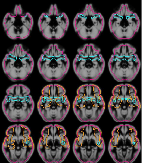
The MCA-M1 was segmented previously on a group of Alzheimer patients: Each segment was normalized to MNI space and the union segment over patients was dilated to construct the MCA-M1 region (Fig 1). Also in MNI space, the vascular territory supplied by the MCA was extracted from an atlas2 (Fig 1). Both regions were coregistered to the mean perfusion weighted image for each of 111 subjects with diabetes II in varying degree, and the AIF was automatically chosen using the k-means based algorithm by Mouridsen et al1.
The number of successfully selected arterial signal curves in MCA-M1, MCA-territory and whole-brain regions were compared based on i) High minimum signal intensity: the number of curves staying above noise level (determined as the signal baseline variation) and ii) Early signal: the number of curves peaking prior to the average WM and GM curves.
Results
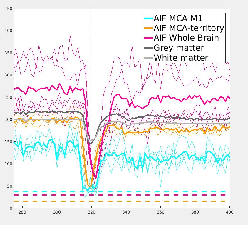
When
selected in the M1 region, the signal
variation increased and the signal peak dropped below noise-level (Fig. 2, blue curve). When selected in the MCA-territory,
the AIF (Fig. 2.yellow curve) did not always peak earlier than the average grey matter
curve (dark gray).The AIF selected from whole brain often peaked even later than gray and white matter (Fig. 2. pink curve).
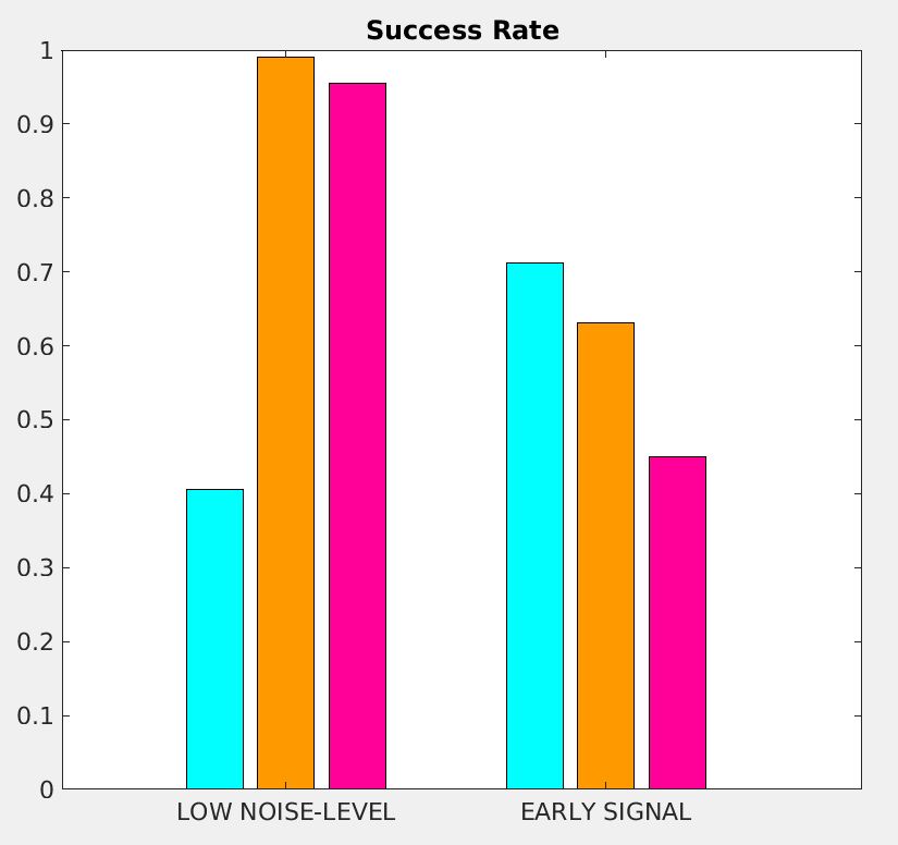
In Fig. 3, the
bar plot shows that less than half of the AIFs were well above noise level when selected in the M1 region. AIFs were successfully selected in the MCA-territory above noise level and with a similar amount of early-peaking AIFs as the AIFs form the M1 region. The whole-brain AIF had a low success rate: in less than half of the patients, the AIF peaked earlier than the tissue curves.
Discussion
AIF selection in or around the M1 segment of MCA is
problematic due to high risk of signal truncation. The increased noise level will introduce larger variance in the estimated perfusion parameters as shown by Erbinger et al3. Without spatial restriction on the search algorithm (whole brain), AIFs with venous contamination were selected, and these curves were delayed compared to gray matter. This would lead to underestimation of the mean transit time.
This study demonstrates that the MCA vascular territory is a better target allowing for robust automatic AIF selection without introducing significant delays. In a new approach, the search region can be reduced to only voxels where the signal peaks prior to the average GM and WM curves and where the signal drop is below noise level.
Acknowledgements
No acknowledgement found.References
Arterial territories of the human brain: cerebral hemispheres.Tatu L, Moulin T, Bogousslavsky J, Duvernoy H.Neurology. 1998 Jun;50(6):1699-708
Automatic selection of arterial input function using cluster analysis.Mouridsen K, Christensen S, Gyldensted L, Ostergaard L.Magn Reson Med. 2006 Mar;55(3):524-31
Reliable perfusion maps in stroke MRI using arterial input functions derived from distal middle cerebral artery branches.Ebinger M, Brunecker P, Jungehülsing GJ, Malzahn U, Kunze C, Endres M, Fiebach JB.Stroke. 2010 Jan;41(1):95-101
Figures