2171
Reconstructing Pseudo-Continuous Arterial Spin Labeling Perfusion Signals through Modulation of Labeling RF Power and Fourier Analysis1Department of Bio and Brain Engineering, Korea Advanced Institute of Science and Technology, Daejeon, Republic of Korea, 2Gordon Imaging Center, Massachusetts General Hospital, Harvard Medical School, Boston, MA, United States, 3Department of Radiology, Seoul National University College of Medicine, Seoul, Republic of Korea
Synopsis
The conventional pCASL is vulnerable to data corruption and has high specific absorption rate. In this study, we propose a new pCASL approach using modulation of labeling RF pulse power and Fourier analysis. The proposed approach enabled us to acquire perfusion images comparable to those of the conventional pCASL. Under data corruption, the proposed approach maintained the perfusion signals well with no observable effect, while the conventional method showed almost no perfusion signal. The proposed approach has relatively low average SAR and instantaneous SAR, potentially advantageous at high fields. These advantages of the proposed method warrant further investigation.
Introduction
Pseudo-continuous arterial spin labeling (pCASL) is the perfusion imaging technique recommended by the ASL community [1]. Perfusion signals are mostly acquired by subtraction of images from label and control scans. In conventional pCASL, labeling and control scans are performed by pseudo-continuous short RF pulses with phase cycling of 0° and 180°, respectively. Recently, a new pCASL strategy, which does not use any RF pulse for control scan, has been suggested [2]. The approach provides higher sensitivity, robustness to scan conditions, and reduced specific absorption rate (SAR) than those of the conventional pCASL. Also, pair-wise subtraction typically used in ASL is vulnerable to unintended image corruption, which may be overcome by period modulation of labeled signals and corresponding Fourier analysis. In the new pCASL (no-RF‑control pCASL), the difference between labeling and control scans is the RF power (flip angle), and thus it is possible to modulate the labeling effects by changing the RF power. In this study, we propose a new approach for pCASL by periodic modulation of labeling RF pulse flip angle and Fourier analysis of the modulated data (with no separate labeling or control scan). The proposed method is expect to have low instantaneous SAR and be robust to unintended image corruption during data acquisition because of the Fourier analysis.Data Acquisition
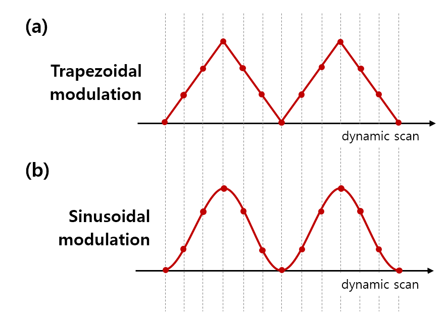
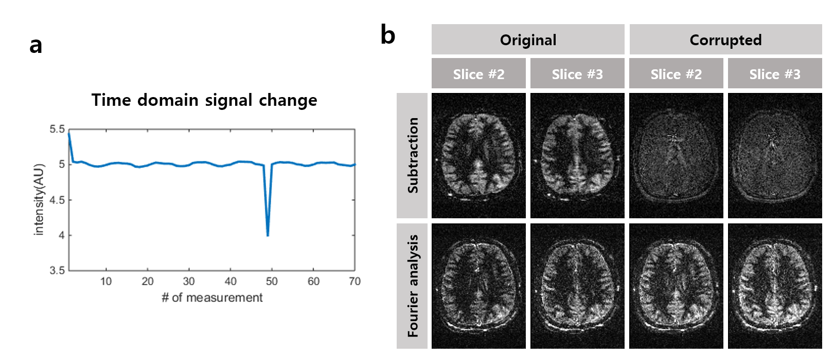
All experiments were performed on a Siemens 3T Trio system (Siemens Medical Solution, Erlangen, Germany) on 3 healthy volunteers. The selection gradient of the pCASL labeling scan increased to 15 mT/m to minimize the MT effects and thus enable no-RF‑control pCASL [2]. Three-dimensional balanced steady state precession (bSSFP) was used for readout. Flip angle of labeling RF pulses of no-RF-control pCASL was changed periodically between 0⁰ and 30⁰ for every dynamic scan with two modulation schemes of trapezoidal and sinusoidal shapes (Fig. 1) and with the step size of 6⁰ and 10⁰ (power-modulated pCASL). Conventional pCASL and no‑RF-control pCASL (with alternating labeling/control scans) were also performed for comparison. Common parameters used in all experiments were as follows; Labeling duration = 1.5 s; labeling RF pulse duration/spacing = 0.5/1.2 ms; TR/TE=4.08/2.04 ms; 70 dynamic scans; readout flip angle = 30⁰. The conventional pCASL was performed with selection gradient of 6 mT/m. Pixel-by-pixel Fourier transform along temporal dimension was performed for power-modulated pCASL data. The location of modulation frequency for each condition was calculated. Also data corruption was simulated by changing intensity of a randomly-selected data by 20% in order to evaluate the robustness of the conventional pCASL subtraction scheme and the proposed method.Results
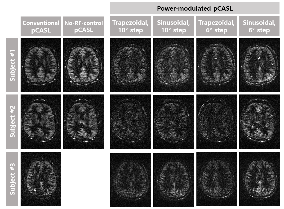
Fig. 2 shows the perfusion image acquired from conventional pCASL, no-RF-control pCASL, and the proposed power-modulated pCASL. The power-modulated pCASL showed perfusion images comparable to those from the conventional pCASL. The 6⁰ step showed better signals than 10⁰ in both trapezoidal and sinusoidal modulations. Images with the sinusoidal modulation showed clearer and higher perfusion signals than those with trapezoidal modulation. Under the condition of the image corruption (Fig. 3a), perfusion images with the subtraction scheme (conventional pCASL) showed significant contamination with no identifiable perfusion signal, whereas those from Fourier analysis (power-modulated pCASL) showed almost no difference before and after the corruption (Fig. 3b).Discussion and Conclusion
The proposed approach utilized modulation of labeling RF pulse power periodically and acquired perfusion images through Fourier analysis. Perfusion images comparable to those of the conventional pCASL were obtained for the proposed method. We could modulate perfusion signals at the desired frequency and Fourier analysis successfully separated the perfusion signal component. Sinusoidal modulation showed better perfusion signals than those of trapezoidal modulation, in agreement with the expectation that Fourier transform of a sinusoidal signal becomes an impulse signal at the frequency of oscillation and thus concentrates energy in the desired frequency component. Labeling RF power modulation, one of the main idea of proposed method, has advantages in the view point of SAR. Average SAR during the whole scan is significantly reduced in the proposed method because of using no-RF control scan. Furthermore, instantaneous SAR in proposed method is expected to be low because the number of scans with maximum labeling RF flip angle (i.e., labeling scan) is reduced. Thus, the proposed method is potentially advantageous at high field MRI systems. Also, Fourier analysis in temporal direction is robust to accidental image corruption. Based on the many advantages demonstrated in this study, it is worthwhile investigating the proposed approach as an alternative pCASL scheme.Acknowledgements
No acknowledgement found.References
1. Alsop D, Detre J, Golay X et al. Recommended implementation of arterial spin-labeled perfusion MRI for clinical applications: A consensus of the ISMRM perfusion study group and the European consortium for ASL in dementia. Magnetic Resonance in Medicine. 2015;73(1):102-116.
2. Han P, Choi S, Park S. Investigation of control scans in pseudo-continuous arterial spin labeling (pCASL): Strategies for improving sensitivity and reliability of pCASL. Magnetic Resonance in Medicine. 2016;78(3):917-929.
Figures