2168
Denoising arterial spin labeling cerebral blood flow images using deep learning-based methodsDanfeng Xie1, Li Bai1, and Ze Wang1,2
1Electrical and Computer Engineering, Temple university, Philadelphia, PA, United States, 2Department of Radiology, Temple university, Philadelphia, PA, United States
Synopsis
In this study, we use Deep Learning-based (DL) method to denoising ASL CBF images. Convolutional neural networks with a “wide” structure, residual learning and batch normalization are utilized as the core of our denoising model. Comparing to non-DL-based methods, the proposed method showed a significant SNR increase as well as partial volume effects improvement. Also, the DL-based method requires less CBF input images, which significantly shorten the acquisition time and reduce the chance of head motion.
Introduction
Cerebral Blood Flow (CBF) can be non-invasively measured with arterial spin labeling (ASL) perfusion MRI but is subject to the low signal-to-noise-ratio(SNR) [1]. Various methods have been proposed to denoise ASL MRI but only provide moderate improvement. Deep learning (DL) is an emerging technique that can learn the most representative signal from data without prior modeling and has shown state-of-the-art performance on natural image denoising [2]. The purpose of this study was to assess the feasibility and efficacy of DL in ASL MRI denoising.Methods
ASL data were acquired from 280 subjects using a pseudo-continuous ASL sequence (40 control/labeled image pairs with labeling time = 1.48 sec, post-labeling delay = 1.5 sec, FOV=22 cm, matrix=64x64, TR/TE=4000/11 ms, 20 slices with a thickness of 5 mm plus 1 mm gap). CBF images were calculated and spatially normalized into the MNI space using ASLtbx [3,4] and SPM12. The deep ASL CBF denoising model was based on 5 layers of Convolutional Neural Networks (CNNs) [5]. CNNs with a “wide” structure, meaning relatively larger filter size (7x7) and more filters (128) was used for two reasons: larger filters can better utilize spatial correlation among neighboring voxels and more filters are able to capture the pixel-level distribution information more effectively [2]. The network was implemented using the Wider Inference Network (WIN) with no signal pooling and no fully connected output layer as often used in regular CNNs. Grey matter (GM) probability map was incorporated as a regularizer because CBF map shows a similar image contrast to that of a grey matter map. To fasten and stabilize model learning, residual learning and batch normalization were both included. The corresponding DL-based ASL denoising model was dubbed as ASLDLD thereafter. To maximally show the benefit of ASLDLD, we took the mean of first 10 CBF images without smoothing (meanCBF-10) as the input image while used the mean of all 40 CBF images (meanCBF-40) with smoothing and adaptive outlier cleaning [4] as the reference. The ASLDLD was trained with data from 240 subjects’ 3D CBF maps (input and reference). The remained 40 subjects were used as test samples.Results
ASLDLD was compared with current non-DL-based denoising methods regarding the SNR of the resultant CBF images. Figure 1 shows the resultant CBF maps from one representative subject. ASLDLD yielded superior performances to non-DL-based methods. Especially, ASLDLD recovered CBF signal in the air-brain boundaries as marked by the green boxes and signal loss due to partial volume effects as labeled by the yellow boxes. Slightly better texture was obtained by ASLDLD too. Figure 2 shows the SNR performance of different methods. SNR was calculated as the ratio between the mean signal of a GM region-of-interest (ROI) and the standard deviation of a white matter ROI. Compared to the non-DL-based methods, ASLDLD showed a 38.6% SNR increase (p=1.14e-4).Discussion
In this study, we showed that DL-based denoising can substantially improve ASL CBF SNR as well as the partial volume effects even only used the mean CBF map of 10 pairs of ASL control/label image acquisitions. In other words, ASLDLD can be utilized to significantly shorten the typical 5-6 mins acquisition time by 75%, which would substantially reduce the chance of head motions, a big confound in ASL imaging.Acknowledgements
No acknowledgement found.References
[1] Detre etc. Magnetic resonance in medicine, 231:37-45, 1992. [2] Liu etc. arXiv:1707.05414 [3] Wang etc. Magnetic resonance imaging, 262:261-269, 2008. [4] Wang etc. Magnetic resonance imaging, 3010:1409-1415, 2012. [5] Krizhevsky etc. Advances in neural information processing systems, 1097-1105, 2012.Figures
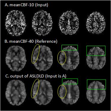
Figure 1: ASL CBF images. From top to
bottom: Input images, non-DL-based denoising output images (reference images), ASLDLD
output images. From left to right: Slices 40, 45, 50 and 55.
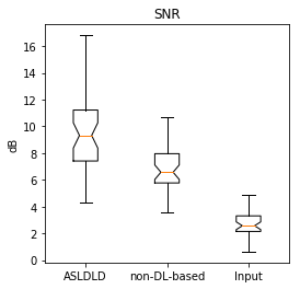
Figure 2: SNR of CBF test images
using different denoising methods.