2163
Tracer kinetics of Velocity Selective Inversion pulses for Arterial Spin Labeling1FMRI laboratory, University of Michigan, Ann Arbor, MI, United States
Synopsis
The tracer kinetic properties of velocity selective inversion pulses were characterized using a two compartment model. The properties of these pulses indicate that VSI pulses can produce large input functions and little or no transit time effects. These translate into speed and SNR gains for perfusion images of both grey and white matter without the use of contrast agents.
Introduction
Velocity Selective Inversion (VSI) pulses have been recently demonstrated as an attractive option for Arterial Spin Labeling (ASL) perfusion imaging because of potential gains in signal to noise ratio (SNR) and decreased sensitivity to bolus arrival time variations. The goal of this work is to characterize the tracer kinetic characteristics of the VSI input function in a 3T scanner using a body coil transmitter in order to facilitate optimal pulse sequence design.Methods
In the first experiment, 13 images were collected with different post inversion delays from 200 to 2600 ms. with no arterial suppression. The experiment was repeated with the arterial suppression pulses turned on.
The spiral acquisition parameters were: matrix size = 128x128x16, voxel size = 0.18x0.18x0.6 cm, BW = 85 kHz, TR = 4000ms., TE = 4.5 ms, nominal flip angle = 30 deg., N. interleaves = 2, N. of images = 10. No VSI or arterial suppression pulses were applied in the first two images of the time series, in order to collect reference images. They were applied in the remaining 8 images alternating between selective and non-selective pulses.
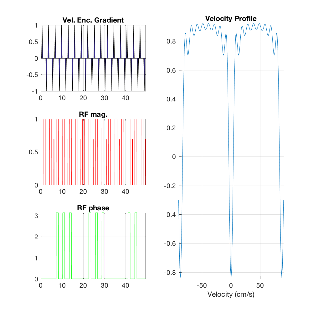
The VSI pulses (figure 1) consisted of a train of 9 segments 20 deg hard pulses (81.25 mG for 160 us) separated by 6 ms gaps. Two refocusing hard pulses (117 mG for 1 ms) were inserted in those gaps with an MLEV phase pattern. Velocity encoding was done along the Z axis by inserting triangular gradient pulses (Gmax=2 G/cm, 300us ramp, 200us gap) of alternating sign between RF pulses. The non-selective case was carried out by using gradient pulses of the same sign. The total duration of the pulse train was 49.4 ms. The arterial suppression pulses were based on the symmetric BIR-8 velocity selective saturation scheme [Guo 2014].
Images were reconstructed, realigned and smoothed with a Gaussian kernel. The ASL signal was calculated as the percentage signal difference between the magnitudes of the selective and non-selective images, relative to the control images (i.e., no prep. pulses). T1 maps were constructed from the non-selective images at each post inversion delay and used to segment grey matter and white matter regions in each subject. Average ASL signals were calculated for white and grey matter separately. This was repeated in both cases (with and without vascular suppression).
A two compartment model (vascular and extra vascular) of the ASL signal was fit to the ASL signal as a function of the post inversion delay. The inversion efficiency in stationary tissue was calculated from the inversion recovery curves and assumed a Rayleigh distribution of arterial blood volumes. Inversion efficiency of the VSI pulse, perfusion rate and arterial blood volume fraction were estimated using a simple grid search algorithm.
Results
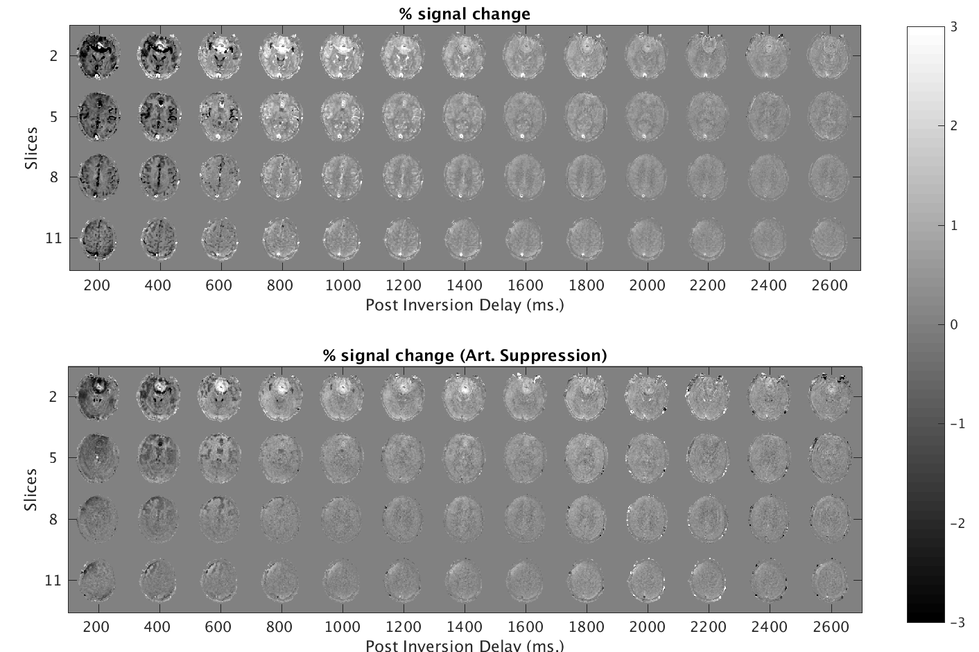
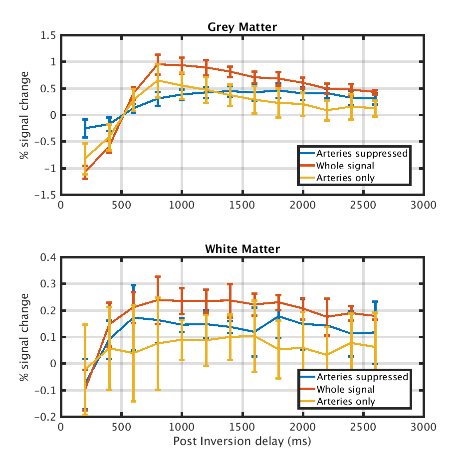
ASL images collected at different times after the VSI pulse can be seen in figure 2. Average ASL time courses over gray and white matter from all participants can be seen in figure 3. Early images after the VSI pulse clearly show the arterial network. This pattern is not apparent when the arterial suppression pulse was used. The sign of the ASL image changes around approximately 800 ms after the inversion pulse. The ASL signal decays afterward. After 1800 ms, the ASL images with and without arterial suppression are indistinguishable.
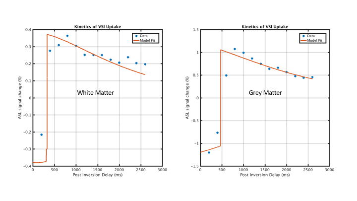
The proposed two compartment model fit to the unsupressed time courses can be seen in figure 4. Fits yielded arterial CBV of 0.90 % and 0.14 % and perfusion rates of 29 .3 and 12.7 ml/min/100g for grey and white matter, respectively.
Discussion
VSI pulses produce perfusion weighted images with high SNR. The kinetics of the VSI input function conforms with the standard two compartment model and can be used to estimate important hemodynamic parameters. No significant bolus arrival delays were observed in the grey or the white matter. The "tail-edge" of VSI arterial input function was not apparent in the explored time delay range (200-2600 ms).
These features translate into speed and SNR gains for perfusion images without the use of contrast agents. VSI pulses are particularly well suited for FMRI and white matter perfusion imaging.
Different input function models and the effects of B1 imperfections on the arterial input function are still under investigation.
Acknowledgements
National Institutes of Health (R21EB021562)
References
Nielsen (2013). Magnetic Resonance in Medicine, 69(2).
Qin (2016). Magnetic Resonance in Medicine, 76(4), 1136–1148
Guo (2014). Magnetic Resonance in Medicine, 73(3), 1085–1094
Figures