2146
Quantitative susceptibility imaging for the assessment of early radiation-induced white matter injury in children with primary brain tumorsJunjie Wu1, Susan Palasis2, Natia Esiashvili3, Richard Jones2, Eduard Schreibmann3, and Deqiang Qiu1
1Department of Radiology and Imaging Sciences, Emory University School of Medicine, Atlanta, GA, United States, 2Department of Radiology, Children's Healthcare of Atlanta, Atlanta, GA, United States, 3Department of Radiation Oncology, Emory University School of Medicine, Atlanta, GA, United States
Synopsis
We examined radiation-induced white matter injury using quantitative susceptibility mapping (QSM) in children with primary brain tumors. Following radiation therapy, susceptibility changed with time and dose. QSM may be a useful biomarker for irradiation damage.
Introduction
Children with brain tumors are commonly treated with whole brain radiation therapy. However, radiation therapy can lead to several adverse effects, including neurocognitive decline. White matter has been reported to be predisposed to radiation-induced injury, which may be a major cause of neurocognitive deficits. QSM is a recently developed MRI method that quantifies tissue magnetic susceptibility from phase images and is sensitive in detecting white matter injury1. In this study, we aimed to evaluate the early changes of radiation-induced white matter injury using the advanced MR technique of QSM.Methods
Five patients with brain tumors (age, 12 ± 3.32 years; 3 females) were included in the study. The patients underwent MR examinations before and after radiotherapy at multiple time points on a Philips Ingenia 3T scanner (Philips Healthcare, Best, the Netherlands) with a 32-channel head array coil. QSM images were acquired using a 3D multi-echo gradient-echo (GRE) sequence: FOV = 230 x 230 mm2, matrix = 256 x 256, slice thickness = 1.5 mm, 108 slices, TR = 35.26 ms, first TE = 7.2 ms, 5 echoes, echo spacing = 6.2 ms. Image processing was performed using a software package developed in-house2. The phase image from each echo was first unwrapped using a Laplacian method followed by spherical mean filtering to remove the background field to generate the filtered field map. The filtered field maps from each echo were then averaged to increase the signal-to-noise ratio. An L1-norm optimization based method was used to reconstruct susceptibility map from the mean field map. QSM images were subtracted by the mean values of cerebrospinal fluid, resulting in normalized susceptibility images. Dosimetry was generated on a CT image and all images were co-registered to correct for differences in positioning. Longitudinal changes of susceptibility were evaluated with respect to regional radiation dose3.Results
Fig 1 shows the iso-dose lines superimposed on the GRE magnitude image of the first echo in a representative patient. Fig 2 displays susceptibility maps before and after radiation therapy in another representative patient. Fig 3 shows mean Δsusceptibility as a function of time following initiation of radiation therapy. Among 3 patients who had a follow-up at approximately 6 months after radiation therapy, increased susceptibility was found within this time window. Mean Δsusceptibility values as a function of dose were shown in Fig 4. In 4 out of 5 patients, increases in susceptibility values were observed. Increase in susceptibility (Δsusceptibility) correlated positively with radiation dose up to 45 Gy, after which a reversal of the trend was observed in 4 out of 5 patients (Fig 4).Discussion and Conclusion
By using QSM, we have successfully detected susceptibility changes in white matter following radiation therapy in children with brain tumors. Susceptibility in white matter increased after radiation therapy as compared to the baseline visit, possibly due to demyelination. QSM therefore could be utilized as a valuable biomarker for radiation-induced white matter damage.Acknowledgements
No acknowledgement found.References
- Wisnieff C, Ramanan S, Olesik J, Gauthier S, Wang Y, Pitt D. Quantitative susceptibility mapping (QSM) of white matter multiple sclerosis lesions: Interpreting positive susceptibility and the presence of iron. Magnetic Resonance in Medicine. 2015;74:564-570.
- Qiu D, Chan GC-F, Chu J, Chan Q, Ha S-Y, Moseley ME, et al. MR quantitative susceptibility imaging for the evaluation of iron loading in the brains of patients with β-thalassemia major. American Journal of Neuroradiology. 2014;35:1085-1090.
- Qiu D, Leung LHT, Kwong DLW, Chan GCF, Khong P-L. Mapping radiation dose distribution on the fractional anisotropy map: Applications in the assessment of treatment-induced white matter injury. NeuroImage. 2006;31:109-115.
Figures
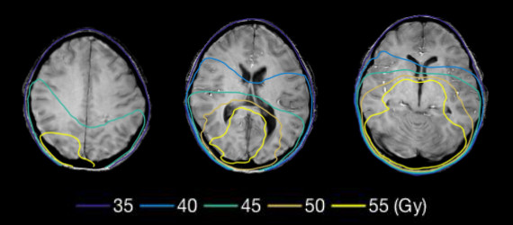
Fig
1. GRE magnitude
image of the first echo in a representative patient, superimposed with iso-dose lines.
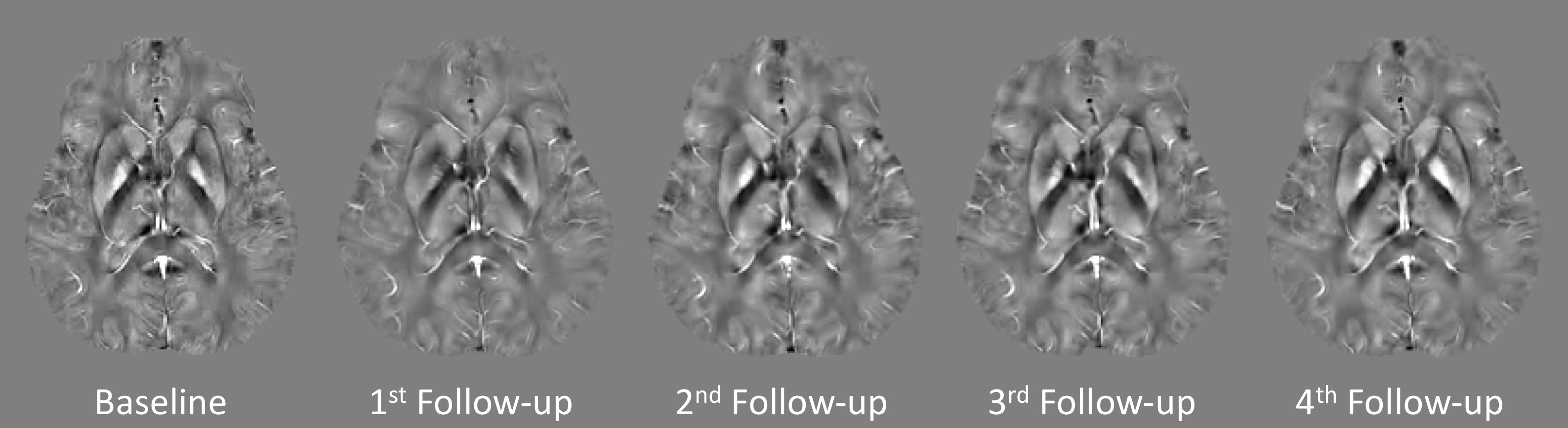
Fig 2. Susceptibility maps at the baseline
visit and follow-up visits after radiation therapy in a representative patient.
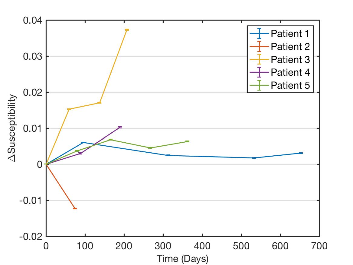
Fig
3. Mean values of Δsusceptibility
as a function of time after radiation therapy. Among 3 patients who had a follow-up at
approximately 6 months after radiation therapy, the mean susceptibility increased within this time
window. Error bars represent standard error of the mean.
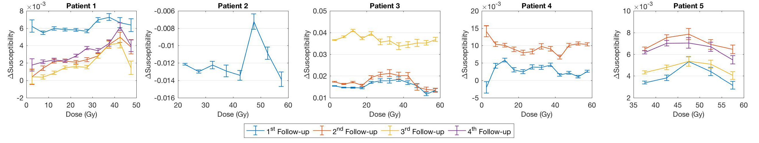
Fig
4. Mean
Δsusceptibility as a function of dose. In 4 out of 5 patients, Δsusceptibility
values were mainly positive. ΔSusceptibility increased with dose, and then
decreased in 4/5 patients. Error bars represent standard error of the mean.