2136
Comparative analysis of diffusion kurtosis imaging, diffusion tensor imaging and diffusion weighted imaging in grading and assessing cellular proliferation of meningioma1Radiology, Fujian medical university affiliated union hospital, Fuzhou, China, 2Fudan university affiliated huashan hospital, Shanghai, China, 3Fujian medical university affiliated union hospital, Fuzhou, China
Synopsis
An accurate evaluation of the WHO grade and cellular proliferation is particularly important in meningiomas, it may facilitate treatment decisions and improve clinical prognosis. But conventional magnetic resonance (MR) imaging were not sufficiently accurate in evaluating the meningioma grade and Ki-67 expression. This study prospectively evaluate and compare diffusion kurtosis imaging (DKI), diffusion tensor imaging(DTI) and diffusion weighted imaging (DWI) metrics in determining the grade and cellular proliferation of meningiomas. It was found that DKI is a better diffusion technique for assessing the grading and cellular proliferation of meningiomas compared to conventional diffusion imaging.
Purpose
Meningiomas are the most common type of intracranial brain tumors.At present, the pre-surgical diagnosis of meningiomas mainly relies on their radiological features. But conventional magnetic resonance (MR) imaging were not sufficiently accurate in evaluating the meningioma grade and Ki-67 expression. This study prospectively evaluate and compare diffusion kurtosis imaging (DKI), diffusion tensor imaging(DTI) and diffusion weighted imaging (DWI) metrics in grading meningiomas, and to assess the correlation between diffusion metrics and the Ki-67 labeling index (Ki-67 LI).Methods
From the Neurosurgical Department of Union Hospital of Fujian Medical University, a total of 107 patients with suspected meningioma on conventional MRI between August 2014 and October 2016 were consecutively enrolled in the study. Preoperative imaging including DWI and DKI was performed, and image data were prospectively analyzed. DKI used a SE-EPI diffusion sequence for image acquisition (TR/TE = 6,000/94 ms, NEX = 1, matrix = 128 × 128, number of sections = 48, sections thickness = 3 mm, spacing = 0 mm, FOV = 24 cm, number of b0 = 3, b values = 1,000 and 2,000 s/mm, number of directions = 30 for each, acquisition time = 6 minutes 24 seconds). All the data were used to calculate the diffusion kurtosis (b = 0, 1000 and 2000s/ mm2) and diffusion tensor (b=0 and 1000) simultaneously. The DKI and DTI data were processed using Diffusional Kurtosis Estimator (version 2.5.1).The diffusion metric maps were processed using Image J (Version 1.50i), including MK, Ka, Kr, MD, FA and ADC. According to conventional MR images, ROIs over the solid tumor and contralateral normal-appearing white matter (NAWM) were semiautomatically delineated using the wand tool in ImageJ. Afterward, same ROIs were taken from anatomic MR images for all parametric maps. Furthermore, the diffusion values in solid tumor were normalized to the corresponding values in the contralateral NAWM to reduce intersubject variation. All values were compared between high-grade and low-grade meningioma by using an independent t-test. Then receiver operating characteristic and multiple logistic regression analysis were used for comparing diagnostic and predictive efficiencies among these metrics. The relationships between diffusion metrics of meningioma and Ki-67 expression were further evaluated.Results
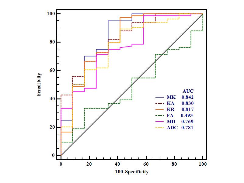
Figure 1shows the manifestations of low and high grade meningiomas respectively on MR images. The normalized DKI metrics(MK, Kr and Ka) were significantly higher in high-grade meningiomas than in low-grade meningiomas (P<0.001), the MD and ADC were significantly lower in high-grade meningiomas than in low-grade meningiomas (P<0.005, 0.007, respectively)(Figure 2). MK(AUC = 0.842) had significantly greater AUC values than MD(AUC = 0.769), and fractional anisotropy (AUC = 0.493) in the differentiation of low-grade and high-grade meningiomas (P= 0.038, 0.002 respectively; Table 2, Figure 3). The multiple logistic regression analysis showed that MK was a significant predictor positively associated with the meningioma grade in the differentiation between HGMs and LGMs (P < 0.001). Significant correlations were found between Ki-67 LI and the kurtosis metrics (P < 0.001), as well as for MD and ADC (P = 0.007, 0.003, respectively).Discussion and conclusion
The MK had greater diagnostic and predictive efficiencies than conventional diffusion parameters in grading meningioma. DKI can characterize non-Gaussian water diffusion, while conventional diffusion imaging techniques only assume a Gaussian distribution of water diffusion. In biologic systems and tumor tissue, the diffusion of water molecules in vivo environment always follow a non-Gaussian distribution. It is reported that kurtosis was more accurate and sensitive for the detection of microstructural changes(3), and MK was able to indicate microstructural complexity in tumor tissue(4). In addition, significant correlations were revealed between Ki-67 LI and the kurtosis metrics, MD and ADC. As such. Kurtosis is believed to be generally proportional to the complexity of the microstructure(5), thus kurtosis metrics is likely to increase while diffusion values are likely to decrease. Meanwhile, the presence of an elevated Ki-67 expression in HG-meningiomas indicates increased mitotic index and enhanced cell proliferation. Consequently, cellular proliferation of meningioma can be non-invasively quantified by diffusion kurtosis metrics. In conclusion, compare to conventional diffusion imaging, DKI is a better diffusion technique for assessing the meningioma grade and cellular proliferation, which have a valuable impact on the diagnosis, treatment decision and prognosis evaluation of meningiomas.Acknowledgements
No acknowledgement found.References
[1] Watanabe Y et. al., EUR J RADIOL. 2013.
[2] Tang Y et. al., AM J ROENTGENOL. 2014.
[3] Lu H et. al., NMR BIOMED. 2006.
[4] Van Cauter S et. al., Radiology. 2012.
[5] Tietze A et. al., AM J NEURORADIOL. 2015.
Figures
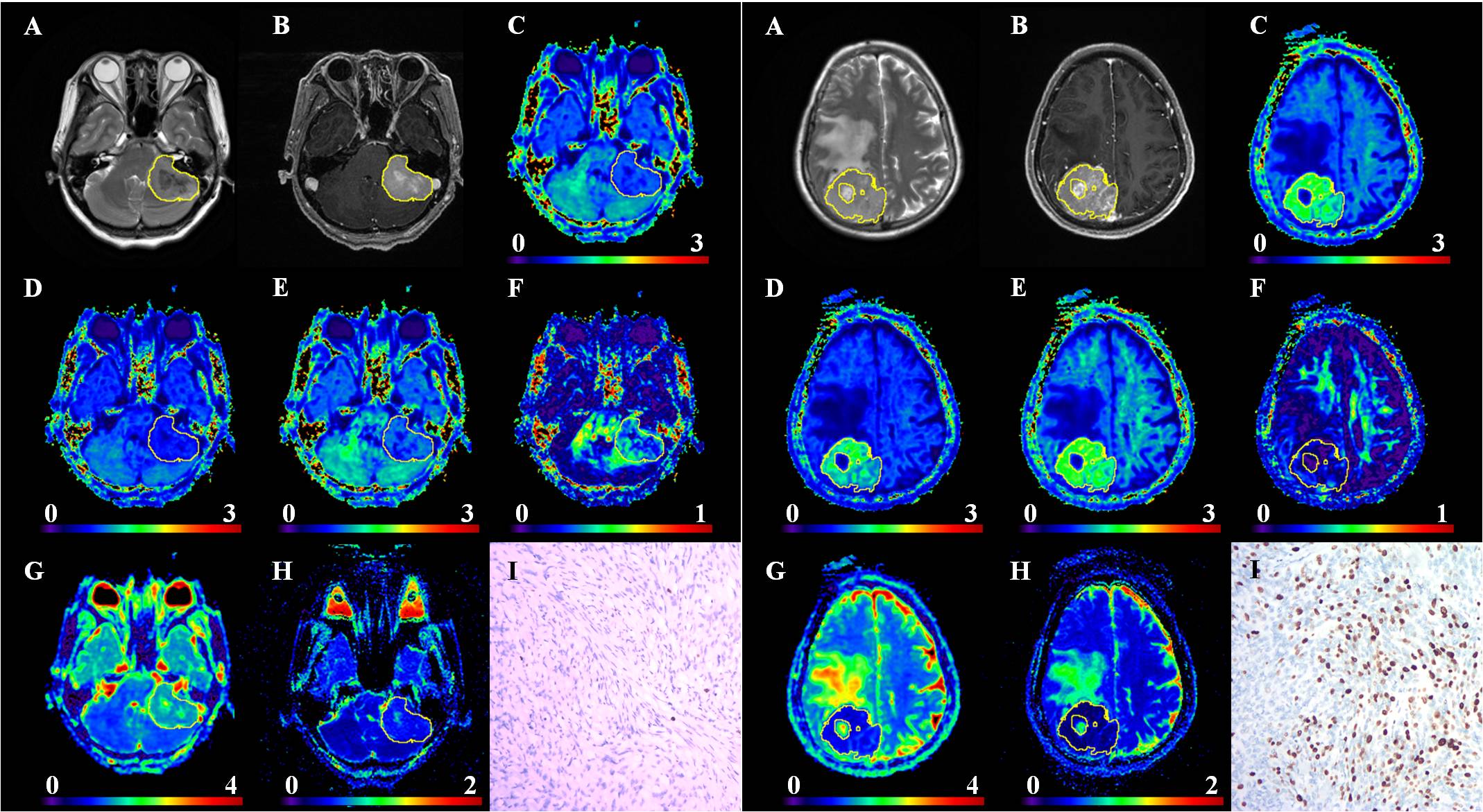
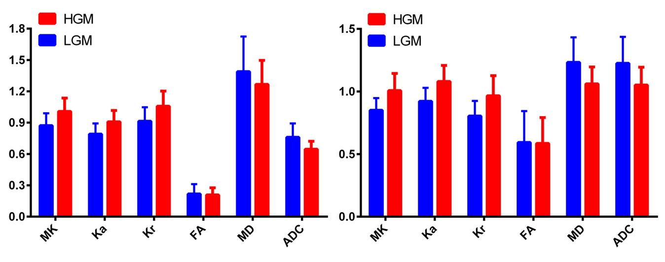
Figure 2: Comparisons of the diffusion metrics between high and low-grade meningiomas.Left: Each diffusion metric in the solid region of the tumor between high and low-grade meningiomas. Right: Parameters in solid tumor normalized to the contralateral NAWM.