2123
Comparison of BOLD and MION enhanced CBV fMRI to the noxious stimulus in anesthetized rhesus monkey1Center for Neuroscience Imaging Research, Suwon, Republic of Korea
Synopsis
Using contrast agent in fMRI has the benefits of providing additional information of regional cerebral blood volume (CBV) and of enhancing sensitivity. Response comparisons of BOLD and MION enhanced CBV fMRI to the noxious stimulus in non-human primate showed signal increase for MION fMRI in the regions that are important in pain-processing network. Activities of some brain areas, including putamen, were captured with MION fMRI, not with BOLD. Capsaicin treatment augmented the responses of fMRI to the noxious stimulation for BOLD and MION fMRI. New insight can be obtained for the pain network through the comparison between BOLD and CBV fMRI.
Introduction
Mapping brain network that is involved in processing the strengths of noxious stimulations with BOLD fMRI in human1 is critical not just in understanding the neural mechanism of pain processes, but in developing treatment of pain. Although there are many advantages, BOLD fMRI has some drawbacks, including nonlinearity2 and low functional sensitivity.3 Because these characteristics of BOLD fMRI can affect adversely for interpreting data, other efforts that can enhance sensitivity would be beneficial in understanding the network of brain.4 MION enhanced CBV fMRI suits well for the purpose, because of its higher sensitivity. Here, we compared responses obtained from BOLD fMRI and MION enhanced CBV to reduce the individual variation, to enhance the contrast-to-noise (CNR) of fMRI and to obtain result from the animal model similar to human network. To understand sensitive network in pain processing, we compared responses to capsaicin, which has been used to model and hyperalgesia in animals5 and humans.6Methods
Animals and experimental procedure Male rhesus macaques (5 yrs, 4-9 kg, n=3) were used in accordance with IACUC guidelines. During imaging, medetomidine (0.015mg/kg/hr) was injected continuously through the saphenous vein and 0.3% of isoflurane was ventilated (Datex-Ohmeda, GE Healthcare, WI), which cocktail drugs were used to prevent the cerebrovascular dilation by high dose of isoflurane. Physiologies were controlled and maintained within a normal range throughout the experiments. Capsaicin was prepared by dissolving 1 mg/mL capsaicin in 70% ethanol and 0.1ml solution was applied to the skin underneath the electrode. In some sessions, MION (10 mg/kg) was injected after obtaining BOLD fMRI.
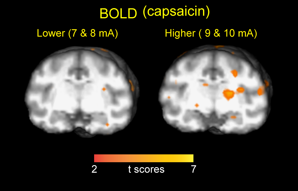
Stimulation Tactile electrical stimulation was applied to the back skin of the lower arm through the attached hydrogel patch electrode (2x2 cm2) without and with topical application of capsaicin. To investigate the intensity dependent fMRI signal, four different strength of electrical stimulation (5Hz, 5sec, 7, 8, 9 and 10mA) was applied in the semi-random inter-trial intervals (29±6.7s). The same stimulation paradigm, including stimulation strength, duration and inter-trial interval, was used for each experimental condition.
Imaging BOLD and MION enhanced CBV fMRI data were collected by a gradient-echo echoplanar whole-brain pulse sequence (EPI; TR=1.2s, TE=22ms, slice thickness = 1.29mm, and 42 slices) using 3T Siemens Prisma. Brain anatomical images (T1; TR=2.3s, TE=3.47ms, slice thickness=0.5mm) of each animal was acquired to co-register with a standard monkey brain atlas for further analyses.
Data analysis Data were analyzed using SPM8 and MATLAB programs. After correcting for head motion, the functional volumes were registered with the anatomical volumes of the animal. Each stimulus epoch was convolved with a canonical hemodynamic response function. Activity maps for each condition were estimated using the GLM. Nuisance covariates were also included to capture noise, including spike and movement-related covariates. Group comparison was conducted using t-test.
Results
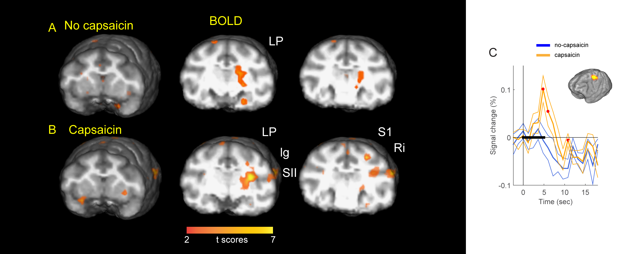
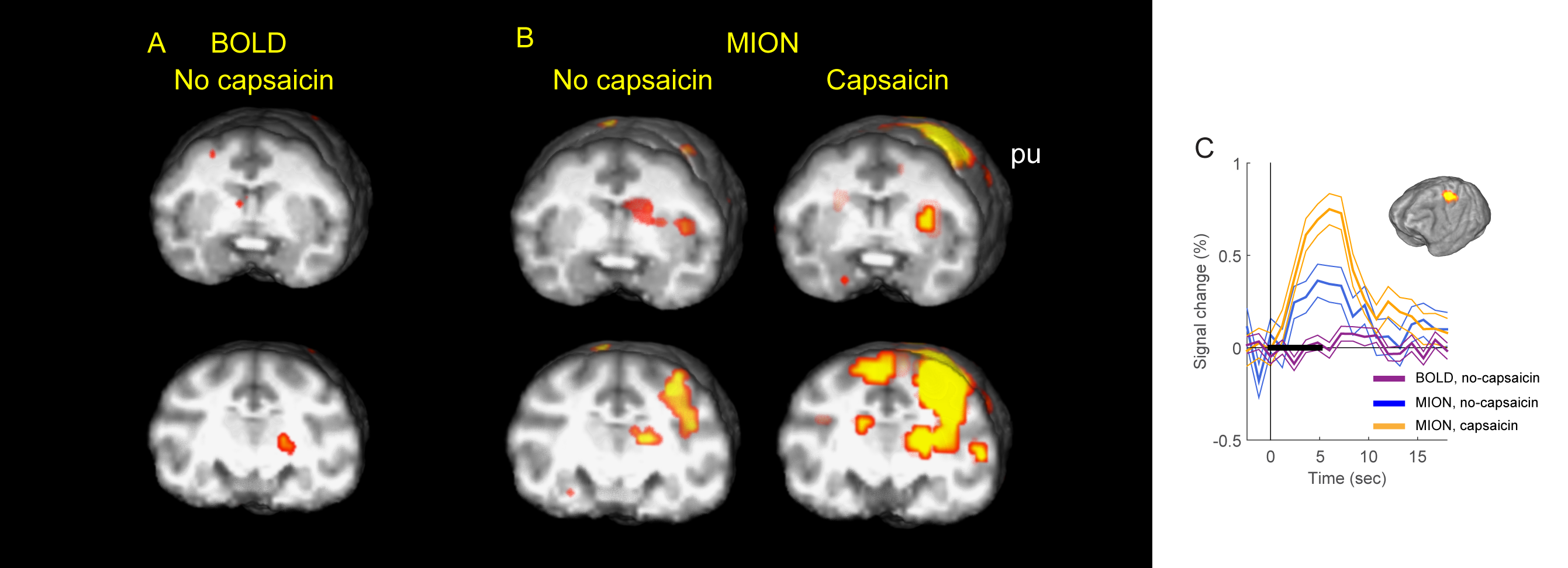
Total seven BOLD sessions for no-capsaicin and capsaicin were obtained from three monkeys. Figure 1A and 1B illustrate responses of BOLD fMRI for each conditions, showing capsaicin potentiated BOLD fMRI responses compared to the no-capsaicin condition in somatosensory, granular insula, secondary somatosensory cortex and retroinsula. Time-course data obtained from ROI of somatosensory cortex (Figure 1C) also confirms different response amplitudes between conditions of capsaicin and no-capsaicin. When trials were divided into two according to the amplitude of stimuli, lower (7mA & 8mA) and higher (9mA & 10mA), many regions, including thalamus and granular insula, showed modulated activation (Figure 2). Figure 3 illustrate results obtained using MION. Significant beta-maps from the same animal were drawn. The use of MION yielded approximately four times larger signal changes relative to BOLD signal at 3T (total n=3). In addition, because of high sensitivity of MION fMRI, subcortical putamen showed activation to the stimulus. Although comparably small, ipsilateral activations of thalamus and somatosensory cortex were also observed in CBV fMRI, which were not obvious with BOLD fMRI.Conclusions
We compared the responses of BOLD and MION enhanced CBV fMRI to noxious stimulation with BOLD and MION enhanced CBV fMRI in non-human primate. Despite the similar map with BOLD fMRI, higher sensitivity in MION fMRI makes it possible to identify clear areal components for pain network and to determine new targets. In addition, mapping brain responses to noxious stimulations in non-human primate and comparing it with human imaging results offer a unique insight for pain processing and gives a clue to the development of an effective clinical target.Acknowledgements
This work was supported by IBS-R015-D1.References
1. Wager TD, Atlas LY, Lindquist MA, Roy M, Woo CW, Kross E. An fMRI-based neurologic signature of physical pain. N Engl J Med. 2013;368(15):1388-97.
2. Gautama T, Mandic DP, Van Hulle MM. Signal nonlinearity in fMRI: A comparison between BOLD and MION. Ieee T Med Imaging. 2003;22(5):636-44.
3. Vanduffel W, Fize D, Mandeville JB, Nelissen K, Van Hecke P, Rosen BR, et al. Visual motion processing investigated using contrast agent-enhanced fMRI in awake behaving monkeys. Neuron. 2001;32(4):565-77.
4. Kim SG, Harel N, Jin T, Kim T, Lee P, Zhao F. Cerebral blood volume MRI with intravascular superparamagnetic iron oxide nanoparticles. NMR Biomed. 2013;26(8):949-62.
5. Asad AB, Seah S, Baumgartner R, Feng D, Jensen A, Manigbas E, et al. Distinct BOLD fMRI Responses of Capsaicin-Induced Thermal Sensation Reveal Pain-Related Brain Activation in Nonhuman Primates. PLoS One. 2016;11(6):e0156805.
6. Iadarola MJ, Berman KF, Zeffiro TA, Byas-Smith MG, Gracely RH, Max MB, et al. Neural activation during acute capsaicin-evoked pain and allodynia assessed with PET. Brain. 1998;121 ( Pt 5):931-47.
Figures