2113
Differences in resting state functional networks during pregnancy in C57Bl6 mice1Experimental 7T MRI Unit, IDIBAPS, Barcelona, Spain, 2CIBER de Bioingeniería, Biomateriales y Nanomedicina (CIBER-BBN) Group of Biomedical Imaging of the University of Barcelona, Barcelona, Spain, 3Neuronal Control of Metabolism (NeuCoMe) Laboratory, IDIBAPS, Barcelona, Spain
Synopsis
The purpose of this study was to investigate if resting state functional MRI is able to reveal brain network changes associated to pregnancy in C57Bl6 mice. 12 mice were scanned before and 3 weeks after pregnancy using a classical resting state fMRI proptocol. Dual regression was performed using these 20 components to find the subject-specific time-series and spatial maps for each network. Significant differences were observed in the striatal, the insula-amygdala and the hippocampal-brainstem networks. Our results reveal that in pregnant C57Bl6 female micethere is reorganization of brain connectivity in specific brain regions and networks.
Introduction
Pregnancy involves radical hormone surges and biological adaptations including short-term and long-lasting changes in the brain1. C57Bl6 mice are widely used as animal models of multiple brain diseases and used as genotypic background for multiple knockout and transgenic strains. The purpose of this study was to investigate if resting state functional MRI is able to reveal brain changes associated to pregnancy in C57Bl6 mice so it can be used as a screening method to further investigate the altered regions at a molecular level.Methods
12 mice were scanned before and 3 weeks after pregnancy on a 7T Bruker BioSpec under medetomide anesthesia (bolus injection of 0.6 mg/kg) and using a single-shot gradient-echo EPI sequence. 420 volumes of 96x96x18 voxels and 0.21x0.21x0.5 mm³/voxel were acquired with TR = 2 s and TE = 10.75 ms. Image preprocessing included: slice-timing, motion correction, skull-stripping, spatial normalization, spatial smoothing, detrending and regression by motion parameters, and temporal filtering (0.01 - 0.1 Hz). 20 independent components were obtained using FSL MELODIC 2 considering the whole cohort. Dual regression was performed using these 20 components to find the subject-specific time-series and spatial maps for each network. The standard deviation of the time-series (Amplitude) of each component and the mean of the Z-values in the spatial maps where each network was localized (Shape Variability) 3 were computed for each subject and the differences between groups were evaluated using Kruskall-Wallis test.Results
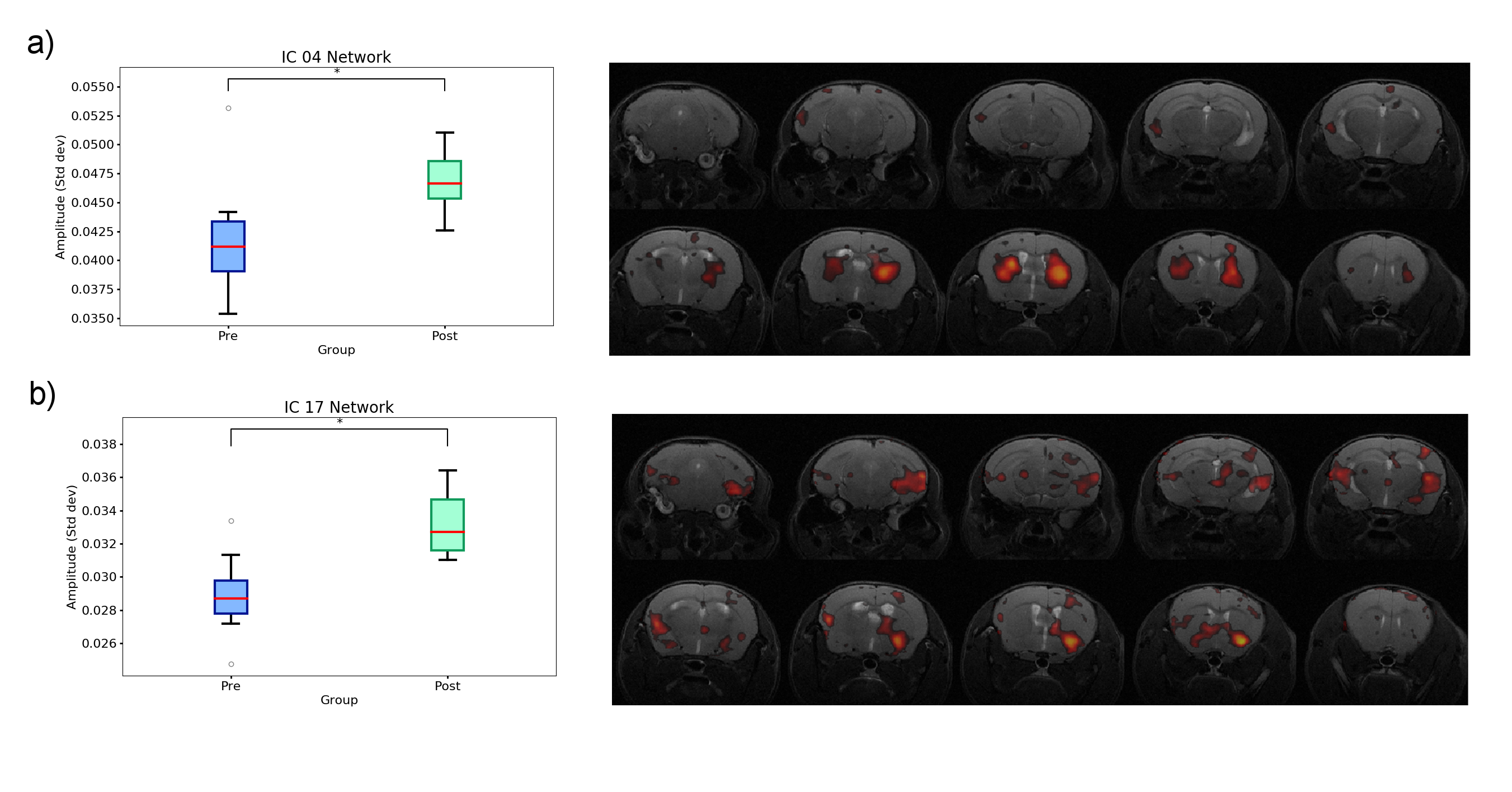
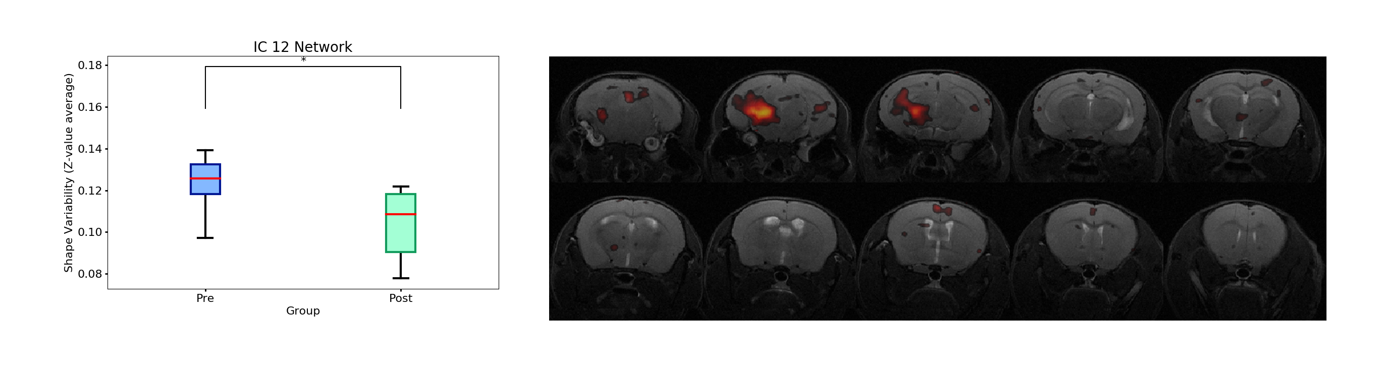
Amplitude was significantly (uncorrected p<0.05) higher in the pregnancy group compared to the pre-pregnant animals in the striatal network (Fig. 1a) and the insula-amygdala network (Fig. 1b). On the other hand, a significant decrease in shape variability was observed in the hippocampal-brainstem network (Fig. 2) in the pregnant females compared to the non-pregnancy group.Discussion
Both the striatal network and the insula-amygdala network have been related to the limbic system. The striatum is more involved in reward and motor responses while the insular cortex and the amygdala have been involved in emotional awareness 4, gustatory functions 5 and in the effective managing of social interactions and the maintenance of social relationships 6. On the other hand, the connectivity between hippocampus and deep mesencephalic nuclei might be related with sleep regulation and memory consolidation systems 7. Therefore, our results reveal that also in experimental animal models such as pregnant C57Bl6 female mice, there is reorganization of brain connectivity in specific regions and networks which are also reorganized in pregnant women 1.Conclusion
Resting state fMRI in mice is a feasible tool to study brain reorganization during pregnancy in experimental animal models and to investigate physiological alterations occurring during this period.Acknowledgements
This work has been funded by the project PI14/00595, integrated in the Plan Nacional I+D+I and co-funded by ISCIII-Subdirección General de Evaluación and European Regional Development Fund (ERDF); and by the Fundació La Marató de TV3 (201441 10). CIBER-BBN is an initiative financed by the Instituto de Salud Carlos III with assistance from the European Regional Development Fund. We are indebted to the Experimental MRI 7T Unit of IDIBAPS.References
1. Hoekzema, E., Barba-Müller, E., Pozzobon, C., Picado, M., Lucco, F., García-García, D., Soliva, J.C., Tobeña, A., Desco, M., Crone, E.A., Ballesteros, A., Carmona, S., Vilarroya, O. Pregnancy leads to long-lasting changes in human brain structure. Nat. Neurosci. 2017; 20:296.
2. Beckmann, C.F., Smith, S.M. Tensorial extensions of independent component analysis for multisubject FMRI analysis. NeuroImage 2005; 25:294-311
3. Nickerson, L.D., Smith, S.M., Öngür, D., Beckmann, C.F. Using Dual Regression to Investigate Network Shape and Amplitude in Functional Connectivity Analyses. Front. Neurosci. 2017; 11:115.
4. Gu, X., Hof, P.R., Friston,K.J., Fan, J. Anterior Insular Cortex and Emotional Awareness. J. Comp. Neurol. 2013; 521:3371.
5. Deen, B., Pitskel, N.B., Pelphrey, K.A. Three Systems of Insular Functional Connectivity Identified with Cluster Analysis. Cereb. Cortex. 2011; 21:1498.
6. Bickart, K.C., Dickerson, B.C., Barrett, L.F. The amygdala as a hub in brain networks that support social life. Neuropsychologia. 2014; 63:248.
7. Picchioni, D., Duyn, J.H., Horovitz, S.G. Sleep and the functional connectome. Neuroimage. 2013; 80:387.
Figures