2106

Pharmacological MRI response of raclopride in rat: relationship with D2 receptor occupancy or cataleptic behavior
Yukiko Masaki1, Yuto Kashiwagi1, Takemi Rokugawa1, and Kohji Abe1

1SHIONOGI & CO., LTD., Osaka, Japan

Synopsis

Pharmacological MRI allows the visualization of brain pharmacological effects of drugs using fMRI. In order to clarify the relationship between fMRI signal and receptor occupancy or behavioral response, 
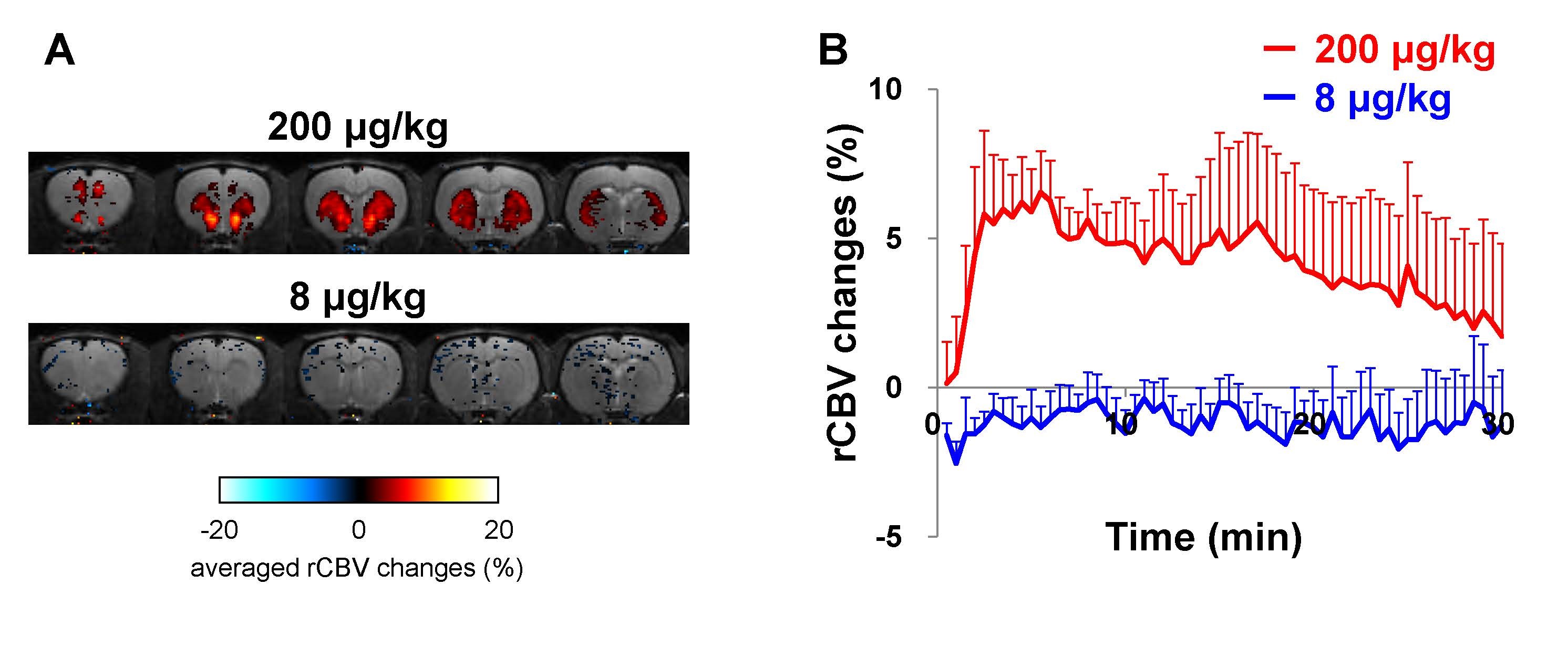
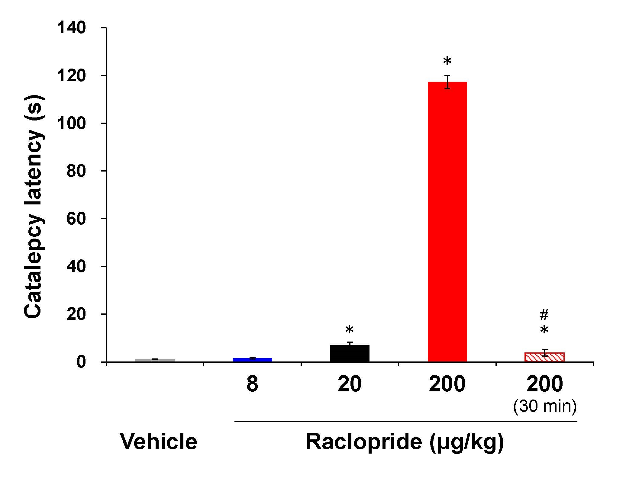
we performed [11C]-raclopride PET, fMRI and the behavioral assessment with raclopride, dopamine D2 receptor antagonist. The positive fMRI response and cataleptic behavior were observed at the dose of raclopride showing 83% of D2 receptor occupancy, but not at the dose of raclopride showing 42% of D2 receptor occupancy. These results suggest that fMRI and behavioral response induced by raclopride will be needed the high D2 receptor occupancy.

Introduction: Pharmacological magnetic resonance imaging (phMRI) allows the visualization of brain pharmacological effects of drugs using functional MRI (fMRI) in clinical and preclinical researches. phMRI is expected to help us facilitate central nervous system (CNS) drug development1. However, there have been few studies demonstrating neurochemical and behavioral response based on the changes in fMRI signal induced by CNS drugs. Therefore, in this study, we performed the successive positron emission tomography (PET) with [11C]-raclopride, fMRI with contrast agent and the behavioral assessment after administration of raclopride, dopamine D2 receptor antagonist to clarify the relationship between fMRI signal and receptor occupancy or behavioral response. Methods: Male Wistar rats (6-7 weeks old) were used in this study. phMRI experiments were conducted at 7T MRI, Varian MRI system 7T/210 (Agilent Technologies) equipped with decoupled volume transmission coil and surface receiver coil (RAPID Biomedical). During the fMRI experiment, blood pressure and heart rate were monitored. Artificial ventilation was used to keep respiration. D-tubocurarine was used to immobilize rats. Relative cerebral blood volume (rCBV)-fMRI were acquired using a multi-slice gradient-echo sequence with imaging parameters2: TR=468.75 ms, TE=10 ms, imaging matrix=128 × 64 (zero filled to 128 × 128), number of averages=1, flip angle=20 degree, and temporal resolution of 30 s. After 10 reference images had been recorded, 20 mg Fe/kg of the blood pool contrast agent Molday ION (Bio PAL) was administered so that subsequent signal changes would reflect alterations in rCBV. Following an equilibration period of 25 min, raclopride (8 and 200 μg/kg) was intravenously administered. The MRI data were acquired over a period of at least 30 min following administration of raclopride. Receptor occupancy was measured at a small-animal PET/CT scanner, Pre-Clinical Imaging System LabPET-12 (Gamma Medica, Inc.) after intravenous administration of a mixture of [11C]-raclopride (20-40 MBq) and non-labeled raclopride (8, 20 and 200 μg/kg). For behavioral study, cataleptic effects of raclopride were measured using a bar-hang test. In the bar-hang test, both forelegs of a rat were gently placed over a 10-cm-high horizontal bar (diameter: 0.5 cm) at 5 min after raclopride administration (8, 20 and 200 μg/kg, i.v.) and 30 min after raclopride administration (200 μg/kg, i.v.), and the retention time in this imposed posture was considered to define catalepsy time. Results: In fMRI study, significant positive fMRI response to raclopride was observed at 200 μg/kg specifically in striatum and nucleus accumbens related to dopaminergic system. fMRI temporal response diminished at 30min after peaking in 5min. In contrast, there was no noticeable fMRI response at 8 μg/kg. PET study showed that receptor occupancy increased dose-dependently (41.8 ± 2.7%, 8 μg/kg; 64.9 ± 2.8%, 20 μg/kg; 83.1 ± 3.0%, 200 μg/kg). Dopamine D2 receptor blockade is known to induce cataleptic behaviors, thus we examined the cataleptic effects by the bar-hang immobility test at 5 min after raclopride administration3. Raclopride at 200 μg/kg significantly increased the catalepsy time compared with vehicle-treated control rats. Raclopride (20 μg/kg) statistically significantly but slightly increased the catalepsy time. At 8 μg/kg, raclopride had not significant effect on the catalepsy time. The catalepsy time at 30 min after raclopride administration (200 μg/kg) substantially decreased compared with that at 5 min after administration. Discussion: The positive fMRI response and cataleptic behavior were observed at the dose of raclopride showing 83% of D2 receptor occupancy, while the dose of raclopride showing 42% of D2 receptor occupancy did not induce fMRI response and cataleptic behavior. These results suggest that fMRI and behavioral response induced by raclopride will be needed the high D2 receptor occupancy. Conclusion: Raclopride-induced fMRI response was observed at doses inducing cataleptic behavior along with high D2 receptor occupancy. phMRI can be useful for drug development as an evaluation method of brain activity which reflects behavioral responses induced by target engagements.

Acknowledgements

No acknowledgement found.

References

1. Jonckers E, Shah D, Hamaide J, et al. The power of using functional fMRI on small rodents to study brain pharmacology and disease. Front. Pharmacol., 2015 Oct 21;6:231 2. Mandeville JB, Marota JJ, Kosofsky BE, et al. Dynamic functional imaging of relative cerebral blood volume during rat forepaw stimulation. Magn Reson Med. 1998 Apr;39(4):615-24 3. Sotoyama H, Zheng Y, Iwakura Y, et al. Pallidal hyperdopaminergic innervation underlying D2 receptor-dependent behavioral deficits in the schizophrenia animal model established by EGF. PLoS One. 2011;6(10):e25831

Figures

Dopamine D2 receptor occupancy in the striatum after administration of raclopride at 8, 20 and 200 μg/kg measured by [11C]-raclopride PET. Data are expressed as means ± SEM (n=3-4).

fMRI responses after administration of raclopride at 8 (n=5) and 200 μg/mg (n=8). A. Mapping of fMRI signals induced by raclopride between 0 and 20 min after administration. B. Temporal profile of raclopride-induced fMRI response in the striatum. Data are plotted as means ± SEM.


Immobility time in the bar-hang test at 5 min after administration of raclopride at 8,20 and 200 μg/kg and 30 min after administration at 200 μg/kg. Data are expressed as means ± SEM (n=6). *p<0.01 vs vehicle. #p<0.01 vs 200 μg/kg at 5min.

Proc. Intl. Soc. Mag. Reson. Med. 26 (2018)
2106