2097
Predicting the age from time of flight MR angiography using 3D convolutional neural network1Seoul St.Mary's Hospital, College of Medicine, The Catholic University of Korea, Seoul, Republic of Korea, 2Yonsei University, Seoul, Republic of Korea
Synopsis
The age-related changes involve the vasculatures of the brain because the brain has rich blood supply. Previous studies using time of flight (TOF) MR angiography suggested that the aging intracranial arteries were tortuous, irregular and heterogeneous in shape. However, the use of these hand-crafted features and qualitative visual assessments are limited in practical clinical use. Vascular aging could be used as an imaging biomarker for the brain if we could distinguish various age-related vascular changes automatically and quickly from MR angiography. In this study, we investigate the feasibility of deep learning based feature extraction as a tool for analysis of age-related change of brain vasculatures.
Purpose
The age-related changes involve the vasculatures of the brain because the brain has rich blood supply.1,2 Previous studies using time of flight MR angiography (TOF-MRA) suggested that the aging intracranial arteries were tortuous, irregular and heterogeneous in shape.3-5 However, since these studies are based on hand-crafted features and visual assessments, they are limited in practical clinical use. Vascular aging could be used as an imaging biomarker for the brain if we could distinguish various age-related vascular changes automatically and quickly from TOF-MRA. In this study, we investigate the feasibility of deep learning based feature extraction as a tool for analysis of age-related change of brain vasculatures.Methods
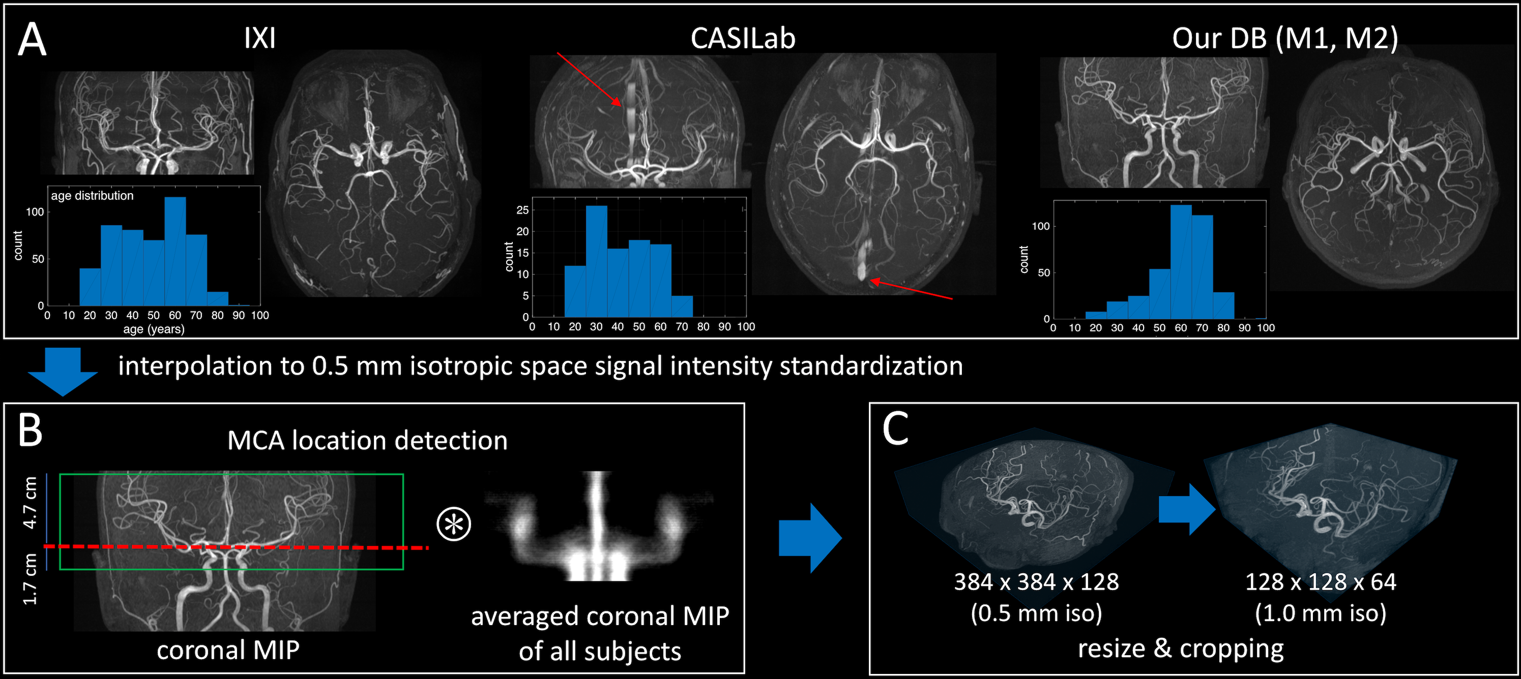
Database A total 950 3D TOF images were collected from two public databases (IXI and CASILab) and our own databases (M1 and M2). Details of the databases are summarized in Figure 1. For IXI and M1 databases, we sampled one in every eight subjects for validation set after sorting the subject by age. Then, the remaining 652 subjects were used for training. CASILab’s and M2 databases were used testing purpose only.
Pre-processing Because databases have different imaging protocols including spatial coverages and venous saturation pulses (red arrows in Figure 2A), pre-processed data were utilized for age estimation. First, 3D TOF images were interpolated to 0.5 mm isotropic space. Second, signal intensities were standardized by subtracting the mean of data and dividing by the standard deviation of data. To match the z-directional coverages of data, an approximate location including middle cerebral artery (MCA) territory in z-axis was estimated by finding maximum 2D correlation coefficient between coronal MIP image of each subject and averaged coronal MIP image of all subjects as described in Figure 2B. After estimating the approximate location of the MCA, a slab of 6.4 cm thickness (from 1.7 cm below to 4.7 cm above) was extracted and utilized for CNNs. For 3D CNN, the central portion of the slab was cropped and the cropped slab was interpolated to 1.0 mm isotropic space due to the limitations of our computing environment. All processes described above were implemented to be fully automatic.
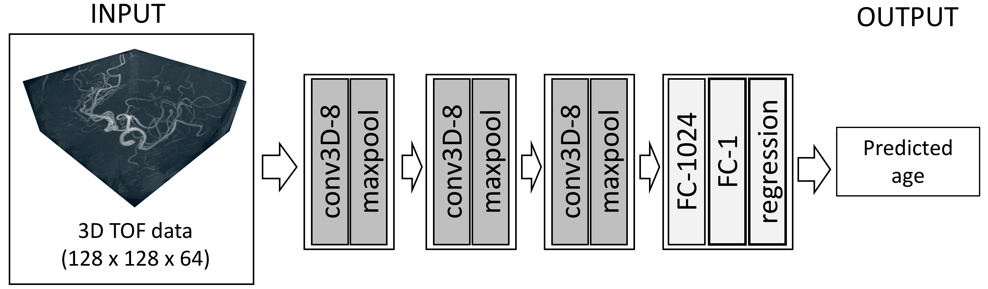
Training CNN A 3D CNN architecture with 5 weight layers (three 3D convolution layers and two fully connected layers) was constructed as described in Figure 3. In this model, 3x3x3 convolution kernels with stride of 1 and 2x2x2 max-pooling with stride of 1 were used for all layers. Training and testing the CNNs were carried out using TensorFlow on a system equipped with a single GPU (NVIDIA, GTX1080).
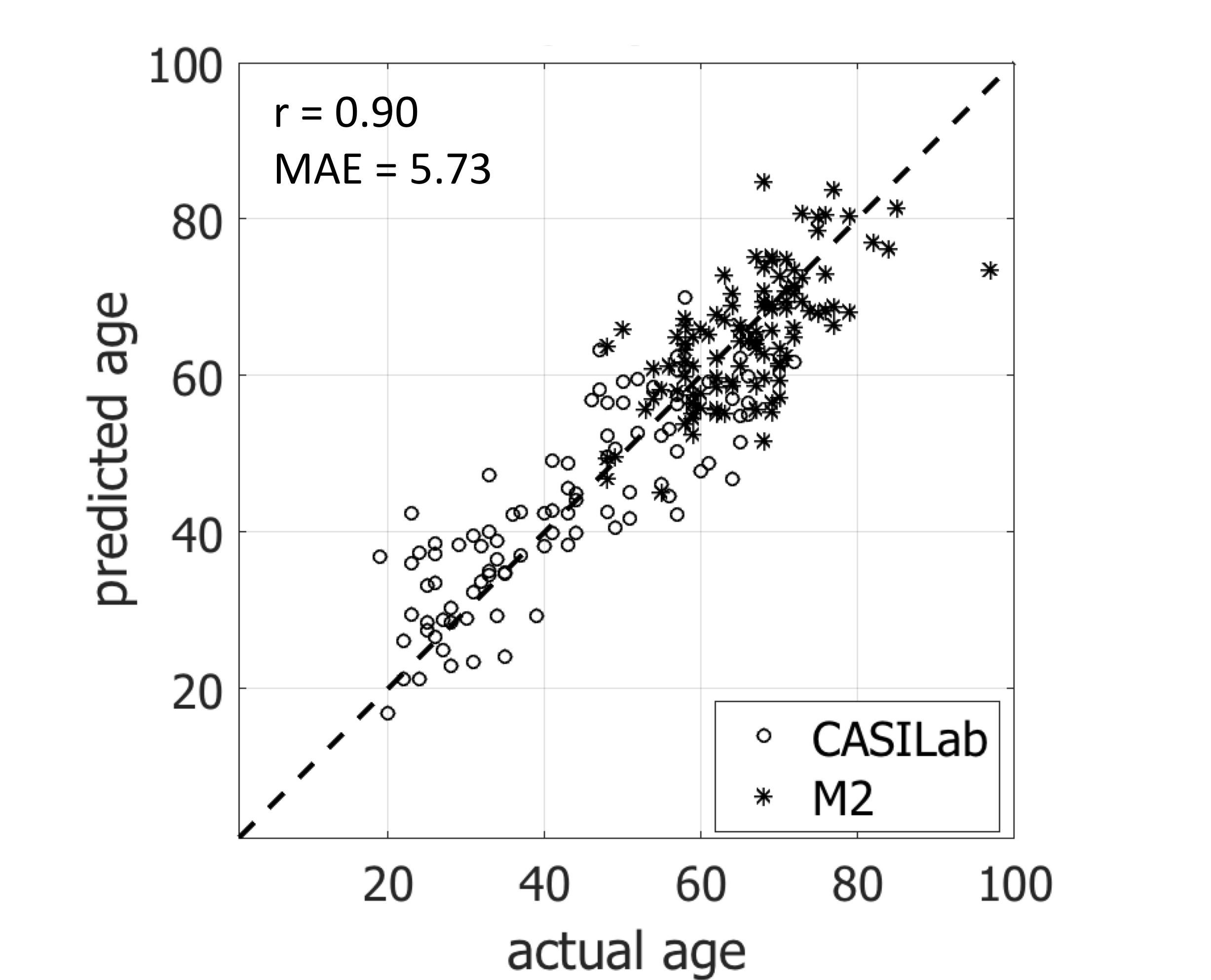
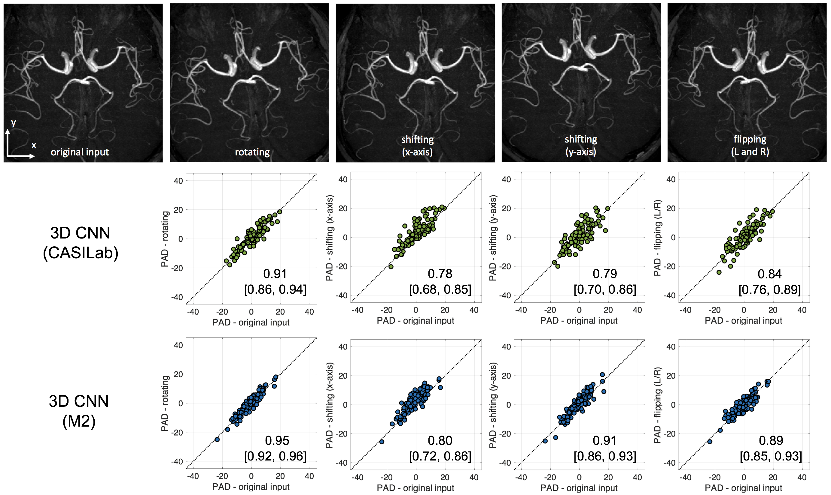
Performance Evaluation The constructed age prediction model from the CNN was evaluated in external test dataset. To evaluate the model accuracy, Pearson’s correlation coefficients and mean absolute errors (MAE) between the actual and predicted ages were calculated. To investigate the sensitivity to the orientation or position of the subject’s head in predicting age, the external test dataset was spatially transformed according to the following four conditions: 1) rotate 7.5 degrees in x-y plane, 2) shift 10 mm in x-axis, 3) shift 10 mm in y-axis, 4) flip left and right. The prediction age differences (PAD) were calculated for each condition and the intra-class correlation coefficient (ICC) was calculated between the PADs from original input data and each condition.
Results
Figure 4 shows the predicted results of test datasets from the trained CNN. The predicted results of test set showed a correlation of r = 0.90 and MAE of 5.73 years for 3D CNN, respectively. For spatially transformed inputs, the PADs from the transformed inputs show a good agreement with the PADS from the original inputs in all conditions as shown in Figure 5. The rotated inputs showed the highest ICCs and the shifted inputs showed relatively low ICCs.Discussion
In this work, we demonstrated the potential of deep learning based CNN model for assessment of aging-related changes from the brain vasculature using TOF-MRA data. In addition, the model showed resistance to spatially transformed input data in predicting ages. For future work, further validations with larger dataset including various conditions will be required. Clinical significance of estimated age using TOF-MRA is also required, which might be used to estimate the accelerated vascular aging.Acknowledgements
The MR TOF images from 486 healthy volunteers used in this study were available from the IXI database (http://brain-development.org/ixi-dataset). The MR TOF images from 94 healthy volunteers used in this study were available by the CASILab at The University of North Carolina at Chapel Hill and were distributed by the MIDAS Data Server at Kitware, Inc. This research was supported by Basic Science Research Program through the National Research Foundation of Korea(NRF) funded by the Ministry of Education (NRF-2017R1D1A1B03033829).References
1. Grinberg LT, Thal DR. Vascular pathology in the aged human brain. Acta neuropathologica. 2010;119(3):277-90.
2. Mrak RE, Griffin ST, Graham DI. Aging-associated changes in human brain. J Neuropathol Exp Neurol. 1997;56(12):1269-75.
3. Bullitt E, Zeng D, Mortamet B, Ghosh A, Aylward SR, Lin W, et al. The effects of healthy aging on intracerebral blood vessels visualized by magnetic resonance angiography. Neurobiology of aging. 2010;31(2):290-300.
4. Kusunoki K, Oka Y, Saito M, Sadamoto K, Sakaki S, Miki H, et al. Changes in visibility of intracranial arteries on MRA with normal ageing. Neuroradiology. 1999;41(11):813-9.
5. Han J, Qiao H, Li X, Li X, He Q, Wang Y, et al. The three-dimensional shape analysis of the M1 segment of the middle cerebral artery using MRA at 3T. Neuroradiology. 2014;56(11):995-1005.
Figures
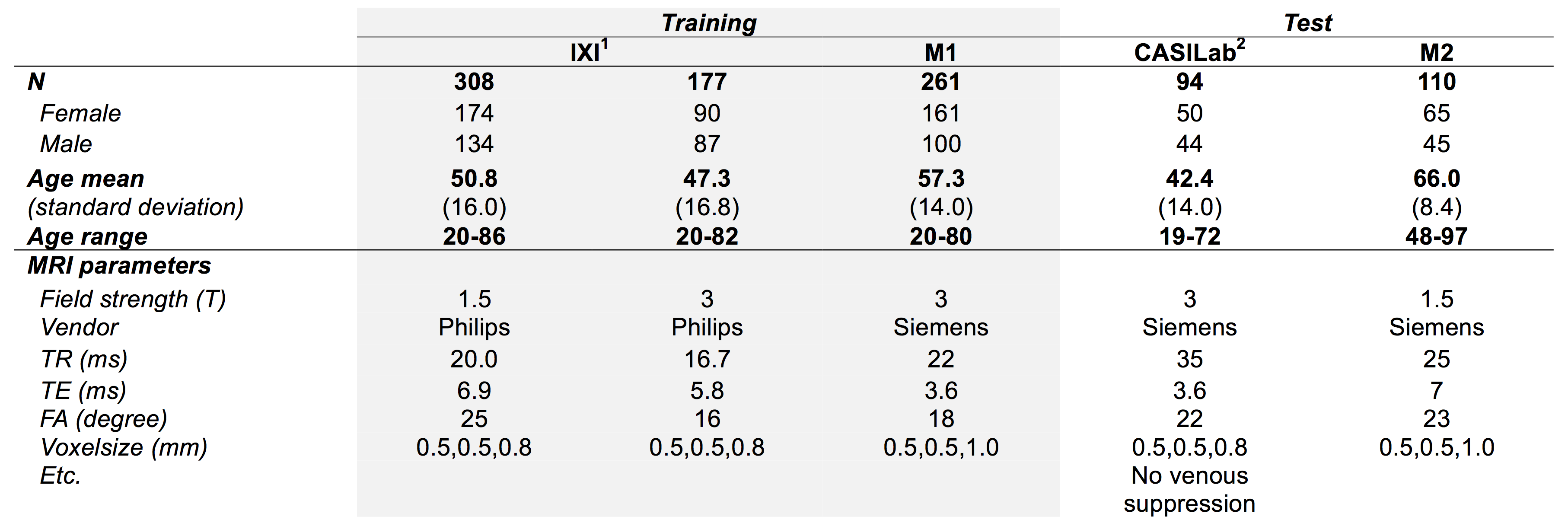
Figure 1. Summary of the databases in the training and test
sets.
1http://brain-development.org/ixi-dataset/
2http://insight-journal.org/midas/community/view/21