2091
Pre-training and training of a Convolutional Neural Network for automatic and accurate hippocampus segmentation from T1-weighted MRI datasets1Medical Sciences, University of Calgary, Calgary, AB, Canada, 2Radiology, University of Calgary, Calgary, AB, Canada, 3Psychiatry, University of Toronto, Toronto, ON, Canada, 4Medicine, Neurology, Sunnybrook Health Sciences Centre, Toronto, ON, Canada, 5Clinical Neurosciences, University of Calgary, Calgary, AB, Canada
Synopsis
The hippocampus atrophy rate (volumetric loss per year) might be a good biomarker for predicting disease progression. However, hippocampus atrophy rate assessment requires accurate delineation of the structure from longitudinal scans. In this work, we propose an automatic approach based on convolutional neural network (CNN) for robust and reliable hippocampus segmentation. Therefore, the CNN was pre-trained using weakly annotated T1-weighted MRI datasets and fine-tuned using fully-annotated datasets. Leave-one-out cross validation revealed that the proposed method leads to robust and reproducible segmentation results with an average Dice coefficient of 0.89.
Introduction
The hippocampus is responsible for episodic memory and learning. Several studies have reported volumetric loss of this structure in Alzheimer’s disease (AD) patients, which is the most common form of late life dementia [1]. The hippocampus atrophy rate (volumetric loss per year) might be a good biomarker for predicting disease progression [2]. However, hippocampus atrophy rate assessment requires accurate delineation of the structure from longitudinal scans. While manual segmentation is tedious, current automatic techniques are often not robust enough to detect small volumetric changes over time [3][4][5][6]. In this work, we propose an automatic approach based on convolutional neural network (CNN) initially proposed by [7][8] for robust and reliable hippocampus segmentation.Methods
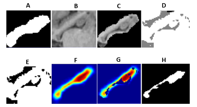
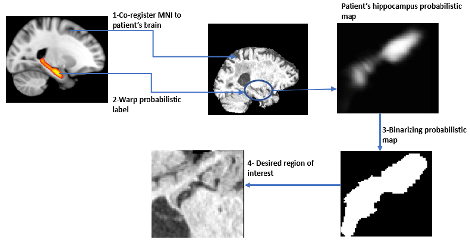
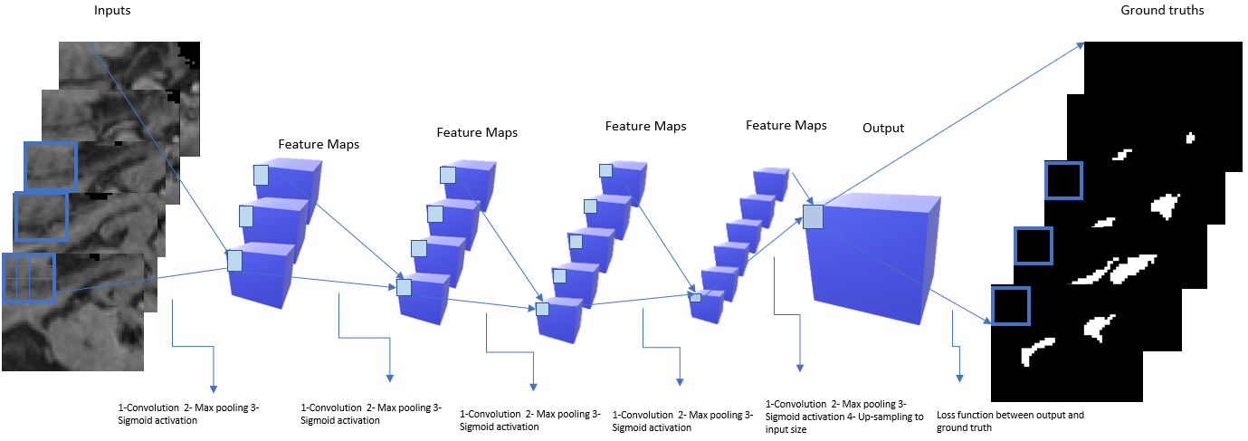
Two different datasets, a weakly annotated and fully annotated dataset, were used for the development and evaluation of the proposed method inspired by [9]. The weakly annotated dataset comprises of 250 T1-weighted MRI images from patients with AD, stroke, mild cognitive impairment, dementia, and healthy subjects. These datasets were weakly annotated (segmented) using a simple automatic approach. Briefly described, this technique performs an atlas-based segmentation of the hippocampus and subsequent post-processing using a k-means clustering approach (Fig. 1). The second dataset used in this work as the fully annotated dataset includes 50 T1-weighted MRI images from patients with AD, mild cognitive impairment, and healthy subjects, which were manually segmented by an expert [10]. The two datasets were pre-processed in the same fashion (Fig. 2) prior to training and testing of the CNN by normalizing the intensity values to zero mean and unary variance values. After this, the MNI brain atlas was non-linearly registered to each dataset using NiftyReg [11]. The probabilistic map of the hippocampus from the Harvard-Oxford subcortical structural atlas was deformed to the patient space using the transformation resulting from non-linear registration and used to define a bounding box encompassing all voxels with a positive hippocampus probability and including an additional small safety margin. Figure 3 shows the architecture of the CNN, which comprises of four convolutional hidden layers subsequent max pooling layer and a sigmoid activation function. The CNN was first pre-trained using the weakly annotated data, whereas the training and testing was restricted to the bounding box in all cases. The main idea of the pre-training is to coarsely optimize the network parameters using a large dataset. After this, the fully annotated datasets were used for fine-tuning of the network parameters in the same fashion.Results
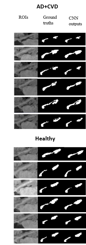
Leave-one-out cross validation using the fully annotated datasets was used for evaluation of the proposed method. The Dice similarity metric was used as a quantitative overlap measurement for segmentation quality assessment. Dice values close to 1.0 indicate a good consensus. Overall, leave-one-out cross validation using pre-training with weakly annotated data followed by fully annotated data resulted in an average Dice coefficient of 0.89±0.015, indicating a very high consensus. Compared to this, training and testing of the CNN using only the fully annotated dataset without any pre-training resulted in an average Dice coefficient of 0.84±0.02, which clearly shows the benefit of pre-training the CNN with weakly annotated datasets. The good quantitative results can also be confirmed visually (Fig. 4). Here, it can be seen that the proposed method is capable of differentiating the hippocampus from other surrounding brain regions such as the amygdala, cerebrospinal fluid, and white matter.Discussion
Overall,
the first results of the proposed hippocampus segmentation method
using a deep convolutional neural network are very promising and in
the top range of previously described results.
The
typical drawback of deep neural networks requiring a large database
for robust training was overcome in this work by using weakly
annotated data for pre-training, prior to fine-tuning the parameters
of the network using fully annotated datasets. Our results clearly
show the benefit of pre-training using weakly annotated datasets.
Another
major benefit of the proposed method is its simplicity as it only
requires an atlas registration and subsequent application of the CNN.
Although training of a CNN can be time-consuming, the application of
a trained network is usually quite fast, especially if only applied
to a small VOI, so that the proposed segmentation method is also
fast. Future research will include further optimization of the CNN
architecture parameters (e.g. number of hidden layers) as well as
validation using test-retest datasets.Conclusion
The proposed technique leads to robust and accurate hippocampus segmentations from patients with various diseases such as AD, stroke, mild cognitive impairment, dementia, as well as healthy individuals and can, therefore, be used in future to determine hippocampus atrophy rates in longitudinal scans.Acknowledgements
No acknowledgement found.References
1. Mu Y, Gage FH. Adult hippocampal neurogenesis and its role in Alzheimer’s disease. Mol Neurodegener. 2011;6(1):85. doi:10.1186/1750-1326-6-85.
2. Carmichael OT, Aizenstein HA, Davis SW, et al. Atlas-based hippocampus segmentation in Alzheimer’s disease and mild cognitive impairment. Neuroimage. 2005;27(4):979-990. doi:10.1016/j.neuroimage.2005.05.005.
3. van der Lijn F, den Heijer T, Breteler MMB, Niessen WJ. Hippocampus segmentation in MR images using atlas registration, voxel classification, and graph cuts. Neuroimage. 2008;43(4):708-720. doi:10.1016/j.neuroimage.2008.07.058.
4. González AM. Segmentation of Brain MRI Structures with Deep Machine Learning. 2012.
5. Lai M. Deep Learning for Medical Image Segmentation. arXiv csLG. 2015;5:2000. http://arxiv.org/abs/1505.02000.
6. Lee N, Laine AF, Klein a, Lee, N., a F. Laine and a K. Towards a Deep Learning Approach to Brain Parcellation. Biomed Imaging From Nano to Macro, 2011 IEEE Int Symp. 2011;(June 2010):321-324. doi:10.1109/ISBI.2011.5872414.
7. Kamnitsas K, Ledig C, Newcombe VF, et al. Efficient multi-scale 3D CNN with fully connected CRF for accurate brain lesion segmentation. Medical Image Analysis. 2017;36:61-78. doi:10.1016/j.media.2016.10.004.
8. Long J, Shelhamer E, Darrell T. Fully convolutional networks for semantic segmentation. 2015IEEE Conference on Computer Vision and Pattern Recognition (CVPR). 2015doi:10.1109/cvpr.2015.7298965.
9. Papandreou G, Chen L-C, Murphy K, Yuille AL. Weakly- and Semi-Supervised Learning of a DCNN for Semantic Image Segmentation. arXiv Prepr. 2015:10. doi:10.1109/ICCV.2015.203.
10. Nestor SM, Gibson E, Gao F-Q, Kiss A, Black SE. A direct morphometric comparison of five labeling protocols for multi-atlas driven automatic segmentation of the hippocampus in Alzheimers disease. NeuroImage. 2013;66:50-70. doi:10.1016/j.neuroimage.2012.10.081.
11. http://cmictig.cs.ucl.ac.uk/wiki/index.php/NiftyReg
Figures