2090
Differences in subcortical brain volumes between expert and novice chess players1Stony Brook University, Stony Brook, NY, United States
Synopsis
The goal of this study was to investigate the anatomical neural correlates underlying expertise acquisition between expert versus novice chess players using MRI. We found that the acquisition of expertise is accompanied by gray-matter volumetric changes in subcortical brain structures implicated in memory and reinforcement learning. By comparison, the anatomical circuits involved in acquired chess expertise differ from other expertise domains. Improving the understanding of the neural correlates underpinning expertise may prove useful in designing individualized training strategies.
Introduction
“Expertise” is the ability to produce high level of performances in a specific domain1. With repeated practice, relevant brain substructures undergo structural and functional changes2 . While MRI volumetric changes in the brains associated with sport and music expertise have been well studied, there have only been a handful of volumetric MRI studies reported associated with chess expertise3,4.
The goal of this study was to investigate the anatomical neural correlates underlying expertise by measuring the brain gray-matter differences between expert versus novice chess players using high-resolution anatomical MRI.
Methods
Data were obtained from an open data set of 29 right handed expert Chinese chess (Xiangqi) players (29±11yo; 9F) and 29 novice chess players (26±7yo; 15F) matched for education level5 . Experts had >2200 ELO rating and training time of 4.24±1.73 hours/day whereas novice group were casual chess players with no significant ELO rating.
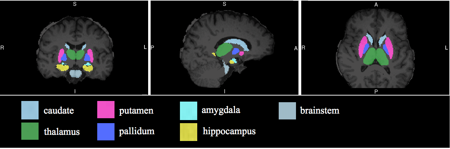
Standard anatomical (1x1x1mm) MRI data were acquired using a 3T MRI scanner. The following major subcortical structures were segmented and analyzed using FSL’s FIRST tool6 : thalamus, caudate nucleus, the putamen, pallidum, hippocampus, amygdala, and nucleus accumben. The left and right hemispheres of these subcortical structures were analyzed separately and tested for lateralization.
Two-tailed unpaired t-tests assuming unequal variance were performed on volumetric fractions between groups. P<0.05 was used as indication of statistical significance adjusted for multiple-comparison. Results were correlated with expertise skill rating.
Results
The results are summarized in Figure 1 and Table 1. The thalamus (left: p=0.0038, right: p=0.0043), the caudate nucleus (left: p=0.00038, right: p=0.00013), the putamen (left: p=0.00016, right: p=0.00032), the pallidum (left: p=0.017, right: p=0.019), and the hippocampus (left: p=0.0081, right: p=0.010) were found to be significantly smaller in the experts as compared to the novices. The total brain volume, however, was not significantly different between groups (p=0.20). No substructures analyzed were significantly lateralized for either the expert or novice subjects. There was no statistically significant correlation in subcortical brain volume with ELO rating after multiple comparison correction.Discussion
The caudate nucleus and putamen are involved in learning and reward function. Duan et al. in 2012 reported smaller caudate nucleus volumes in chess experts compared to non-experts, consistent with our finding3. Our study extended Duan et al.’s study by finding volumetric differences in the putamen, thalamus, pallidum and hippocampus associated with chess expertise. The thalamus is a hub for relaying information from the sensory receptors to proper areas of the brain where it is processed. The pallidum serves as a relay circuit and plays a crucial role in spatial learning. The pallidum, which acquires input from the caudate and putamen and communicates with the subthalamic nucleus, is known to be involved in stimulus discrimination. The hippocampus is involved in memory acquisition, important in expertise acquisition.
There have been many reports on brain volumetric differences associated with sport and musical expertise. Many found MRI brain volumes of specific structures to be larger in experts. Professional golfers (as compared with non-skilled golfers) had larger gray matter volumes in their frontoparietal network, including the premotor and parietal areas7. Professional keyboard players also showed larger gray matter volume in the motor, auditory, and visual-spatial brain regions when compared with a matched group of amateur players and non-keyboard players8,9. A few studies have also reported smaller gray-matter volume in the sports and musical expertise expert groups4,10.
Conclusion
The acquisition of chess expertise is accompanied by gray-matter volumetric changes in subcortical brain structures implicated in memory and reinforcement learning. The smaller brain volume associated with chess experts could be the result of increased neural efficiency. The circuits involved in acquired chess expertise differ markedly from those in sports and music, suggesting functional specialization in the brain is associated with specific expertise domains. Improving understanding of the neural correlates underpinning expertise may prove useful in designing individualized training strategies to achieve the highest performance in the shortest time.Acknowledgements
No acknowledgement found.References
[1] Ericsson, K. A., Charness, N., Feltovich, P. J., & Hoffman, R. R. (Eds.). (2006). The Cambridge Handbook of Expertise and Expert Performance (1 edition). Cambridge ; New York: Cambridge University Press.
[2] Zatorre, R. J., Fields, R. D., & Johansen-Berg, H. (2012). Plasticity in Gray and White. Nature Neuroscience, 15(4), 528–536.
[3] Duan, X., He, S., Liao, W., Liang, D., Qiu, L., Wei, L., … Chen, H. (2012). Reduced caudate volume and enhanced striatal-DMN integration in chess experts. NeuroImage, 60(2), 1280–1286.
[4] Hänggi, J., Koeneke, S., Bezzola, L., & Jäncke, L. (2010). Structural neuroplasticity in the sensorimotor network of professional female ballet dancers. Human Brain Mapping, 31(8), 1196–1206.
[5] Li, K., Jiang, J., Qiu, L., Yang, X., Huang, X., Lui, S., & Gong, Q. (2015). A multimodal MRI dataset of professional chess players. Scientific Data, 2.
[6] Patenaude, B., Smith, S.M., Kennedy, D., and Jenkinson M. A Bayesian Model of Shape and Appearance for Subcortical Brain NeuroImage, 56(3):907-922, 2011.
[7] Jäncke, L. (2009). Music drives brain plasticity. F1000Prime Rep, 1(78). https://doi.org/10.3410/B1-78
[8] Gaser, C., & Schlaug, G. (2003). Brain Structures Differ between Musicians and Non-Musicians. Journal of Neuroscience, 23(27), 9240–9245.
[9] Moore, E., Schaefer, R. S., Bastin, M. E., Roberts, N., & Overy, K. (2014). Can Musical Training Influence Brain Connectivity? Evidence from Diffusion Tensor MRI. Brain Sciences, 4(2), 405–427.
[10] Draganski, B., Gaser, C., Kempermann, G., Kuhn, H. G., Winkler, J., Büchel, C., & May, A. (2006). Temporal and Spatial Dynamics of Brain Structure Changes during Extensive Learning. Journal of Neuroscience, 26(23), 6314–6317.
Figures