2088
Noninvasive Analysis of Brain Shift Transformation in Closed Cranium using MR Images Acquired in Different Body Positions1Information Science and Technology Center, Kobe University, Kobe, Japan, 2Graduate School of System Informatics, Kobe University, Kobe, Japan, 3Department of Neurosurgery, Hyogo Emergency Medical Center, Kobe Red Cross Hospital, Kobe, Japan, 4Department of Radiology, Kobe University, Kobe, Japan, 5Department of Chemical Science and Engineering Faculty of Engineering, Kobe University, Kobe, Japan, 6Department of Neurosurgery, Kobe University, Kobe, Japan
Synopsis
Although the transformation of brain tissue during craniotomy is a well-known phenomenon, there has been a lack of methodological analysis related to physiological brain shift in the closed cranium. In this study, we analyzed brain shift and transformation using MRI volume data acquired in different body positions. The volume data were divided into voxels. Each voxel of the prone or the right volume was registered using voxels of the supine or the left volume as templates, and movement and rotation of each voxel were recorded. Experimental result shows that the displacement in the depth of the brain tended to be conspicuous and rigid compared to the displacement of the brain surface.
Introduction
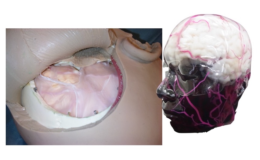
We
are developing a soft material brain model for neurosurgical training1 as shown in Figure 1. In
the process of extracting 3.0T MRI data of brain tissue for application to
additive manufacturing technology, we found that the configuration and position
of the brain changed slightly according to head position. Although the
transformation of brain tissue during craniotomy (so-called brain shift) is a
well-known phenomenon, there has been a lack of methodological analysis related
to physiological brain shift in the closed cranium. In this study, we analyzed
shift and transformation of the brain using MRI volume data acquired in
different body positions.Materials and Methods
Acquire MR images: Three-dimensional MR images of the head of a healthy volunteer (38 years old, male) were acquired using 3.0T MRI with fast spin echo with four different posture. The imaging conditions were as follows: TR/TE, 2500 ms/270 ms; slice thickness, 1 mm; field of view, 256 × 256 mm2; slice gap, 0.5 mm; acquisition matrix, 256 × 256; spatial matrix, 512 × 512 × 400. And patient positions were supine, prone, left lateral decubitus and right lateral decubitus.
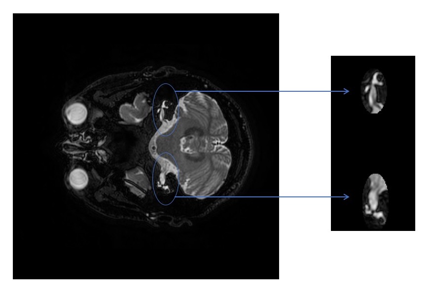
Volume registration: Two step of volume registration of MRI volume data acquired in different position was performed. In first step, whole head registration based on the three-dimensional phase-only correlation method2. The phase only correlation method was a method of finding the correlation between signals focusing on only the phase component obtained by discrete Fourier transform of the signal and was robust against various disturbances such as image brightness change and image quality degradation. It was possible to perform signal alignment and similarity evaluation. In second step, Registration based on anatomical reference organs was performed to the registered volumes by the whole brain registration. We chose left and right internal auditory meatus and the following three semicircular canals as the reference organs because these did not move by posture changes. Intensity base registration was performed with two ellipsoids area includes the reference organs shown in Figure 2.
Analysis of brain shift: The volumes of supine, prone, left-side and right-side were divided into voxels of 20x20x20 pixels. Each voxel of the prone volume or the right volume was registered using voxels of the supine volume or the left volume as templates, and movement and rotation of each voxel were recorded.
Results and Discussion
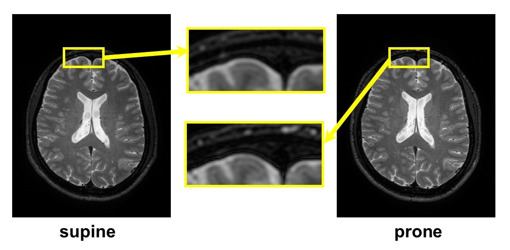
As
shows in Figure 3, The cerebrum was slightly displaced toward the occipital
region or the left side. However, displacements were different for each place.
It was suggested that the cerebrum was deformed.
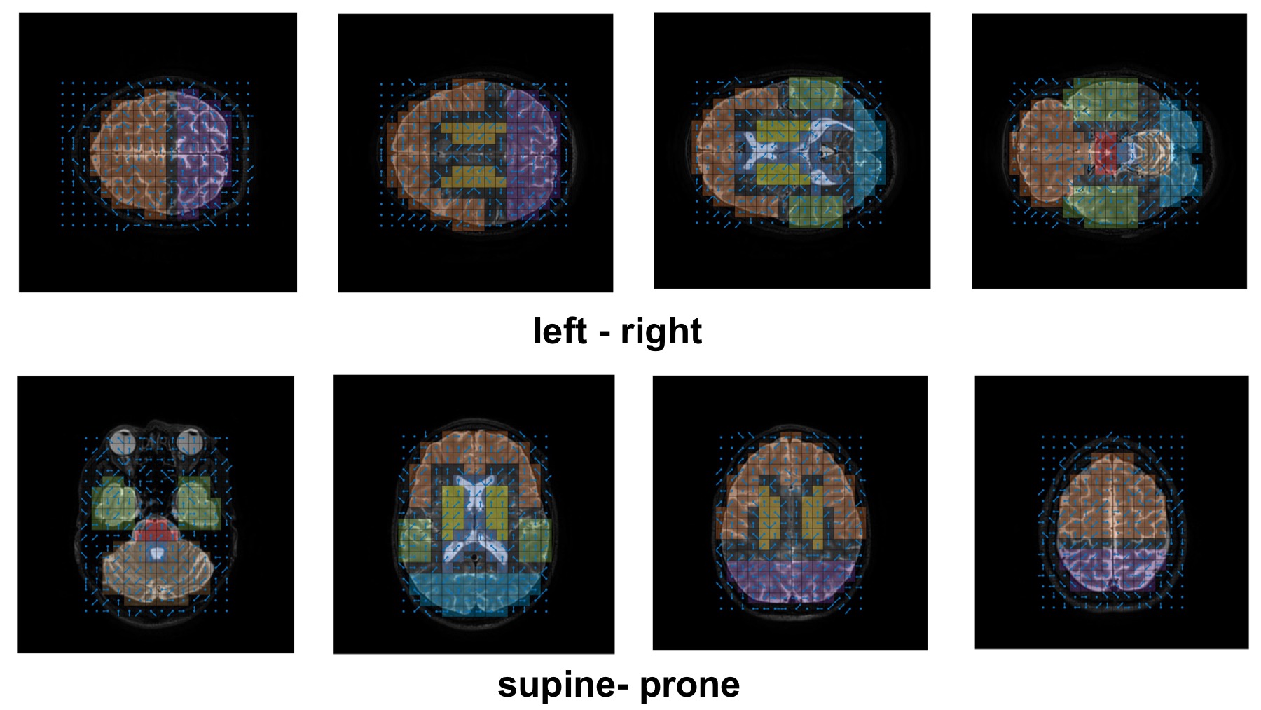
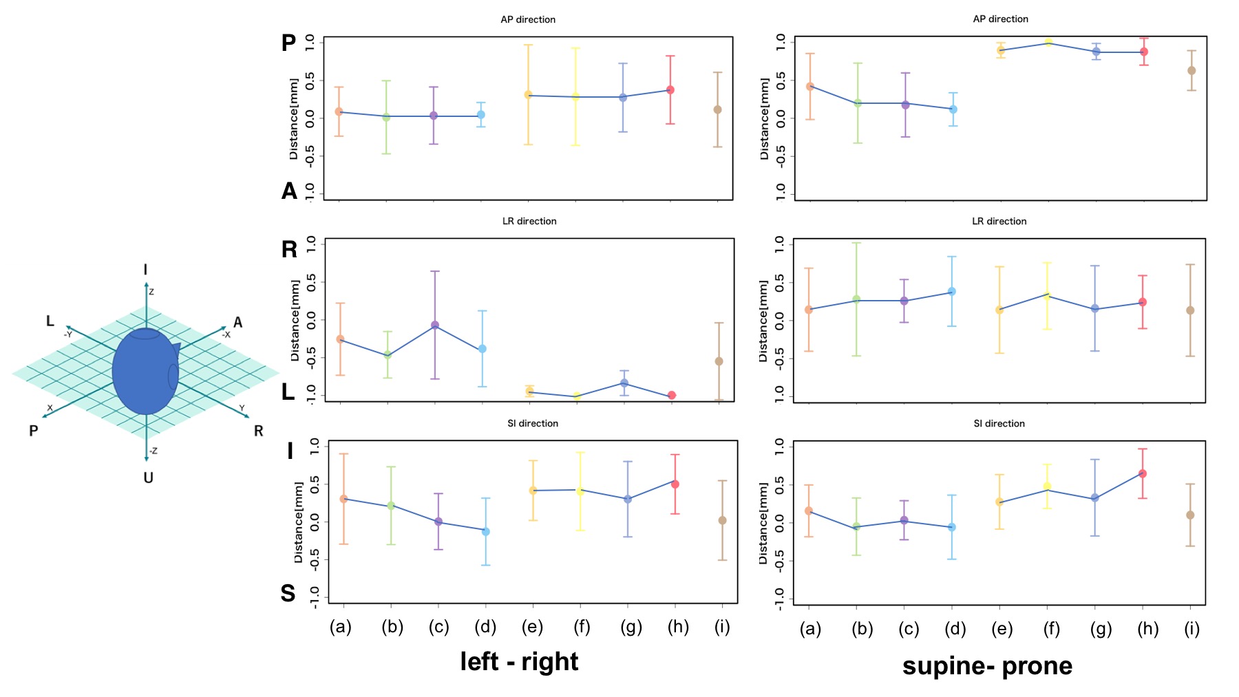
Figure 4 shows the displacement
vector for each voxel in the posture changes of left-right and supine-prone. And
Figure 5 shows the average and variance of the vector component (x, y, z) in each organ. The posture conversion from
the right lateral decubitus position to the left lateral decubitus position
showed displacement of the radial crown, ventricle, basal ganglia, and brain
stem collectively in the rear left downward direction. The movement to the left
was significant. When the posture convert from the prone position to the supine
position, displacement of the radial crown, ventricle, basal ganglia, and brain
stem collectively in the rear right downward direction. The movement to the
downward was significant. Conclusion
The
brain parenchyma was deformed by the body position transformation and displaced
in the cerebrospinal fluid. The factors such as the elastic modulus of the
brain parenchyma to be as soft as 2 kPa and constraints due to the dura
structure are thought to diversify brain movement and deformation. The detail
analysis will be performed using such as optical flow in the future.Acknowledgements
This research is supported by grant number 15K01329 from the JSPS Grant-in-Aid for Scientific Research (C).References
(1) M. Kotera, S. Hayashi, and T. Kubo, Development of Neurosurgical Training Model using Additive Manufacturing Technology, Seikei-Kakou, vol. 27, pp. 359-363, 2015.
(2) Y. Tajima, et al. Fast and Robust 3D Correspondence Matching and Its Application to Volume Registration, IEICE TRANS. INF. & SYS., Vol.96, No.4, pp.826-835, 2013.
(3) Rhoton, Cranial Anatomy and Surgical Approaches, Lippincott Williams & Wilkins, pp187-209, 2007.
Figures