2086
MRI based texture analysis on FLAIR and ADC to predict malignant transformation of Low Grade Gliomas1Radiolgy, Tongji Hospital, Tongji Medical College, HUST, Wuhan, China, 2Radiolgy, Weill Cornell Medical College, NewYork, NY, United States, 3Neurology, Weill Cornell Medical College, NewYork, NY, United States, 4Neurological Surgery, Weill Cornell Medical College, NewYork, NY, United States, 5Biomedical Engineerring, Cornell University, Ithaca, NY, United States
Synopsis
Low grade gliomas (LGG) may undergo malignant transformation into high-grade gliomas, which generally occur within 5 years in about 50% of patients. Hence assessing whether or not a LGG will convert to high grade is of great importance in treatment. In this study, we use texture and histogram analyses on preoperative MRI FLAIR and ADC images to predict malignant transformation from low grade to higher grade glioma, as well as to discriminate between astrocytoma and oligodendroglioma. Based on the receiver operating characteristic (ROC) curves from training data, texture analysis had a higher area under the curve (AUC) value than histogram parameters, and it also more accurately predicted whether LGGs would convert and discriminated between astrocytoma and oligodendroglioma in the testing data. Texture analysis on conventional preoperative FLAIR and ADC images can accurately predict malignant transformation of low grade gliomas, as well as discriminate between astrocytoma and oligodendroglioma.
Background and Purpose: The median survival of patients with low grade gliomas (LGG) is highly variable and ranges between 2 and 20 years, depending on the presence or absence of a 1p19q co-deletion and IDH mutational status. Treatment is necessary to prolong survival and prevent transformation into high-grade gliomas, which generally occur within 5 years in about 50% of patients.1 The purpose of this study is to evaluate the feasibility of texture and histogram analyses, with preoperative MRI FLAIR and ADC images, in (1) predicting malignant transformation from low grade to higher grade glioma and (2) differentiating between astrocytoma and oligodendroglioma.
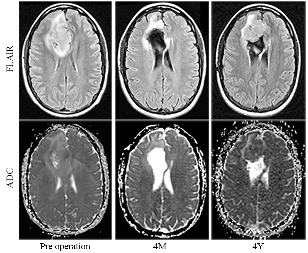
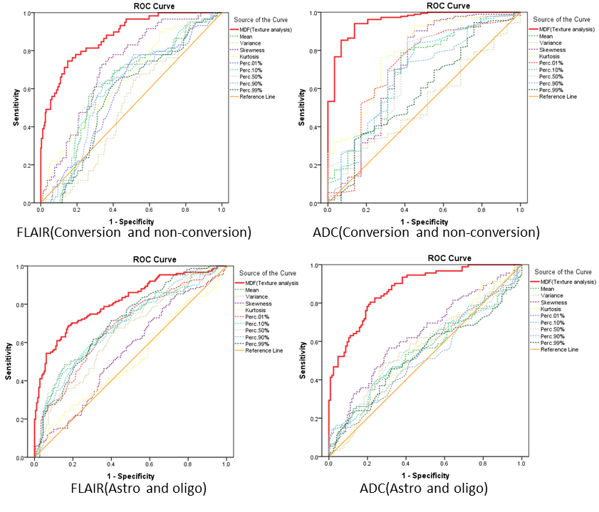
Methods: 14 out of a total of 76 LGGs (36 astrocytomas and 40 oligodendrogliomas) showed malignant transformation to higher grade after 1 to 13 years of follow-up (median 3.5 years). All 14 patients had preoperative FLAIR images and 7 had additional ADC maps. Patients were randomly divided into training (60%) and testing (40%) sets. Regions of interest (ROI) were defined on each slice from the entire tumor volume on FLAIR and ADC images. For texture and histogram analyses, using MaZda software, linear discriminant analysis (LDA) was performed to obtain the most discriminant factor (MDF) values and 9 histogram parameters. Receiver operating characteristic (ROC) curves were utilized in the training group to determine the diagnostic accuracy and obtain the cut-off values for determining the correct rate of discriminating between the two groups in the testing data.
Results: The area under the curve (AUC) using FLAIR and ADC to predict malignant transformation of LGGs was 0.88 (sensitivity 85%, specificity 75%) and 0.96 (sensitivity 86%, specificity 94%). AUC values for texture analysis were greater than those obtained from the 9 histogram parameters (range 0.52-0.72, 0.52-0.78). Using the cutoff value generated to test the testing data, texture analysis correctly predicted malignant transformation in 87% of cases using FLAIR, and 83% of cases using ADC, while the 9 histogram parameters correctly predicted malignant transformation in 19%-81% of cases using FLAIR and 33%-89% of cases using ADC. To discriminate between astrocytoma and oligodendroglioma, the AUC values for FLAIR and ADC were 0.81 and 0.87 respectively, greater than those obtained from histogram parameters (range 0.51-0.71, 0.51-0.63). In the testing data, texture analysis correctly discriminated astrocytoma and oligodendroglioma in 90% of cases using FLAIR and 89% of cases using ADC, while histogram analysis correctly discriminated 43%-60% of cases using FLAIR and 44%-56% of cases using ADC.
Conclusion: Texture analysis on conventional preoperative FLAIR and ADC images accurately predicts malignant transformation of LGGs, as well as discriminates between astrocytoma and oligodendroglioma.
Acknowledgements
No acknowledgement found.References
1. Rotariu D, Gaivas S, Faiyad Z, Haba D, Iliescu B, Poeata I. Malignant transformation of low grade gliomas into glioblastoma a series of 10 cases and review of the literature. Romanian Neurosurgery (2010) XVII 4: 403 – 412Figures