2083
Impacts of Chronic Liver Injury on Brain Energy Metabolism: A 1H-[13C]-NMR Study on Hepatic Encephalopathy1NMR Microimaging and Spectroscopy, Centre for Cellular and Molecular Biology, Hyderabad, India, 2Animal House, Centre for Cellular and Molecular Biology, Hyderabad, India
Synopsis
It has been postulated that excess ammonia and neuroinflammation resulting from liver failure induces astrocytic swelling which can lead to increased BBB permeability and neuronal dysfunction. The impacts of high levels of blood ammonia on the brain energy metabolism is not clear. The objective of current study is to evaluate the neurotransmitter metabolism in CCl4 induced liver injury mouse model using using 1H-[13C]-NMR spectroscopy together with [1,6-13C2]glucose infusion. Our findings indicate reduction in the activity of glutamatergic and GABAergic neurons in the chronic liver damage condition.
Introduction
Liver being a vital organ, controls several metabolic process. The damage to liver impairs its function that creates the metabolic imbalance in the body. Hepatic encephalopathy is a most common disorder that constitutes a spectrum of neuropsychiatric abnormalities, including cognitive deficits seen in patients with liver dysfunction1. Liver play major role in detoxification and removal of ammonia from the blood. Chronic liver insult leads to enhanced ammonia levels in the blood that leads to major metabolic dysfunction. Clearance of excess ammonia from the body is regulated by its conversion to glutamine by combining it with amino acid glutamate. It has been postulated that excess ammonia and neuroinflammation resulting from liver failure induces astrocyte swelling that lead to increased BBB permeability to some molecules, and neuronal dysfunction2. Carbon tetrachloride (CCl4) is an established hepatotoxin which damages liver initially by getting converted into CCl3OO* free radical by cytochrome P450, and later by eliciting myriad of inflammatory signal. The objective of the current study was to understand the impact of liver injury on neurometabolism using Carbon tetra chloride model of hepatotoxicity3.Materials and Methods
All animals protocol was approved by institutional animal ethics committee of CCMB. Male C57BL/6J mice of two months age were divided into two groups: Group I). Olive oil +Carbon tetrachloride (CCl4), Group II). Olive oil. Mice were injected 6% CCl4 dissolved in Olive oil (0.25ml/kg) for twice a week for 10 weeks. The control mice receive the vehicle only. The SGOT (Serum Glutamic Oxaloacetic Transaminase) and SGPT (Serum Glutamic-Pyruvic Transaminase) were measured periodically. After last dose, Elevated Plus Maze(EPM) and Open Field Test (OFT) were preformed to assess anxiety and depression like phenotype. For metabolic analysis, mice were infused with [1,6-13C2]glucose4,5 in awake condition, blood was collected from retro- orbital sinus for analysis of plasma glucose concentration and 13C enrichment. The brain Metabolism was arrested at 7 minutes by using Focused Beam Microwave Irradiation (4.0 kW, for 1.0 s)6. Brain was dissected, and metabolites were extracted as using previous protocol with slight modification7. The concentration and 13C labeling of cerebral amino acids in the tissue extracts were measured ex vivo in 1H-[13C]-NMR spectrum recorded using Bruker 600MHz AVANCE III HD spectrometer8.Student's t test was carried out to find the statistical significance amoung the different groups.The value presented in mean±SD.Results and Discussion
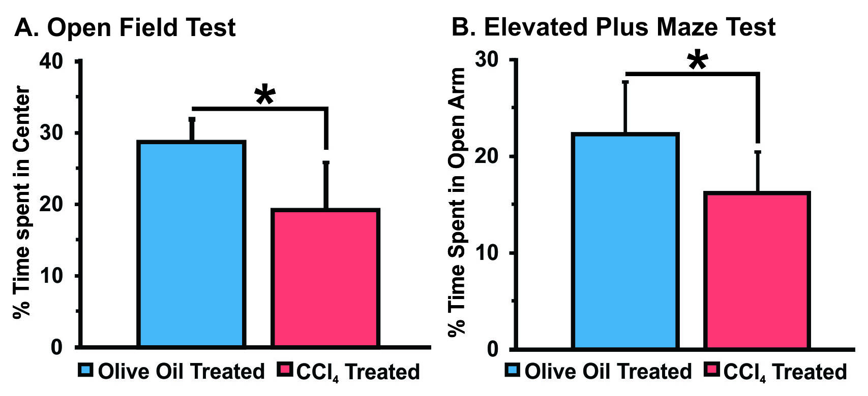
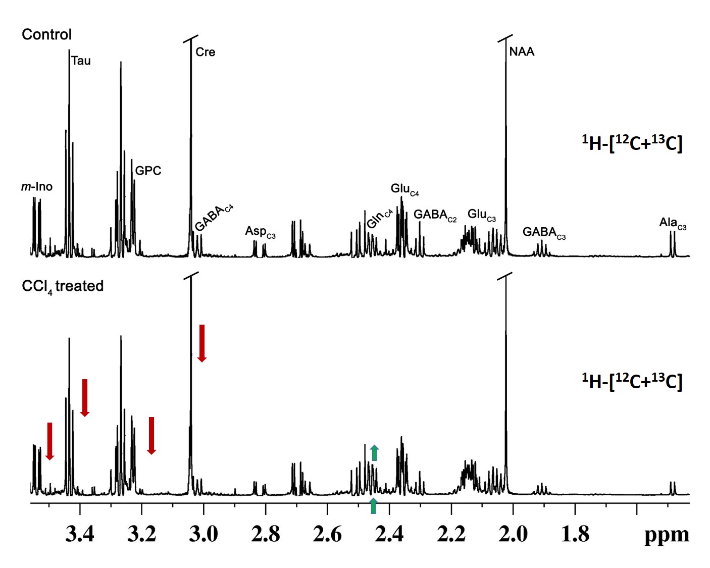
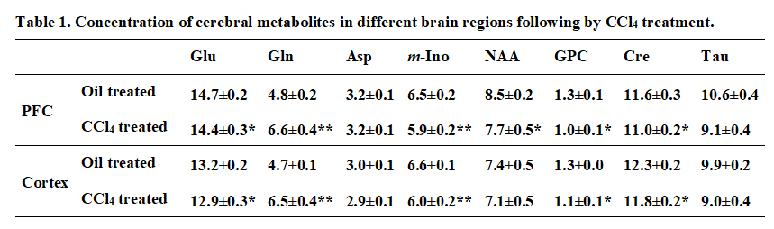
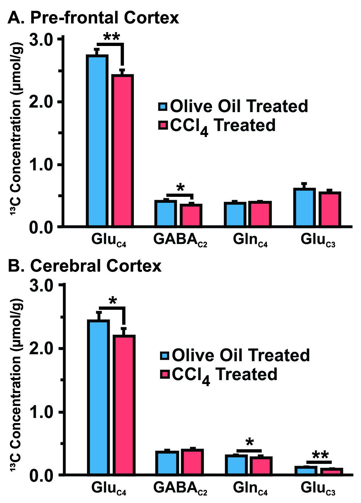
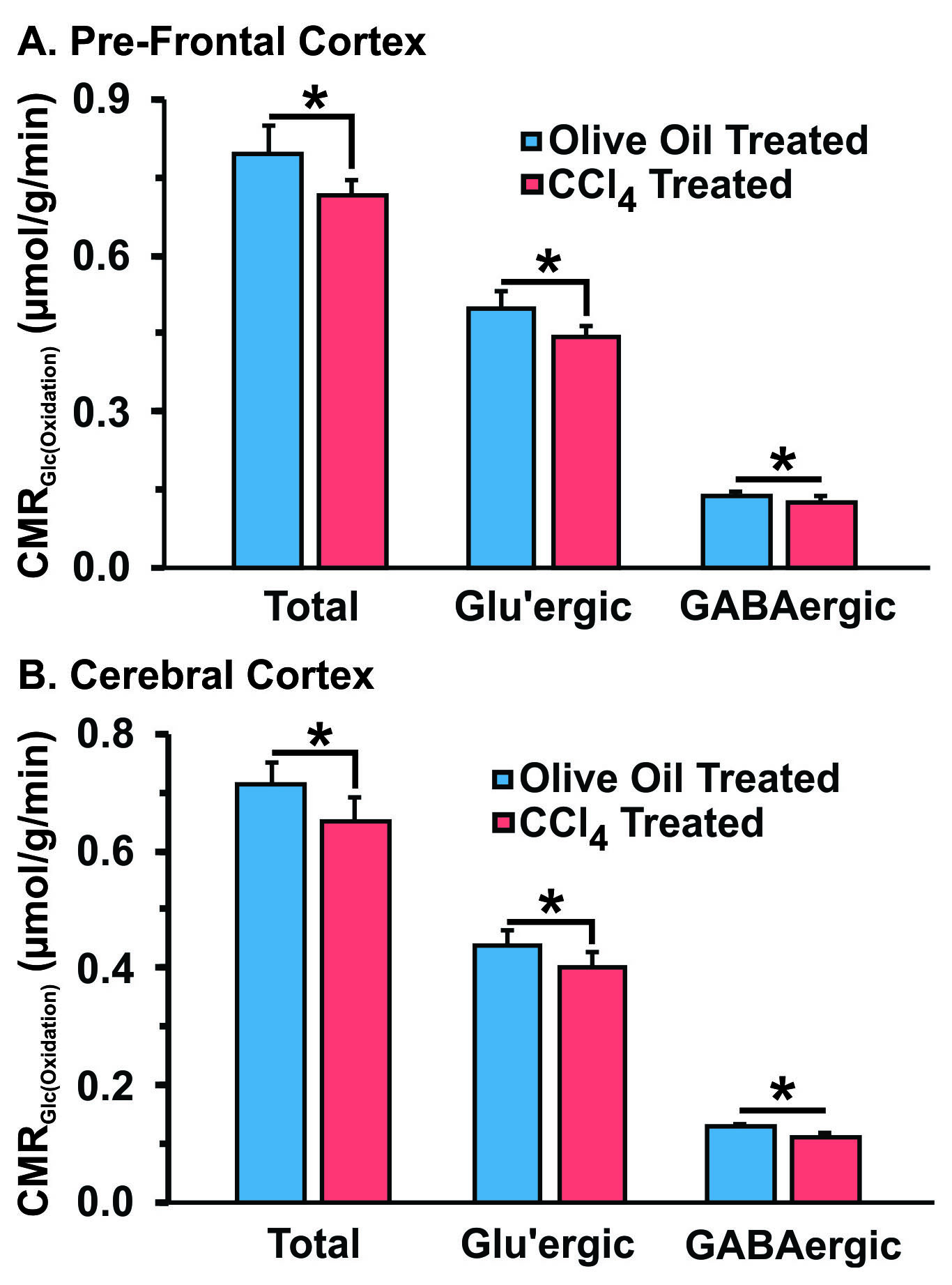
The elevated level of SGOT (CCl4 1106.32±209.8 IU/L; Control 79.2±20.98 IU/L) and SGPT (CCl4 1098.3±200.92 IU/L; Control 28.3±4.2 IU/L) in serum indicated severe liver damage in CCl4 treated mice. The CCl4 treated mice exhibits increased anxiety in the OFT and EPM tests as compared with Olive oil treated Controls (Figure 1) suggesting depression like phenotype. The level of glutamine was elevated significantly (p<0.01) in CCl4 treated mice (Figure 2), and decreased in glutamate, N-acetyl aspartate (NAA), myo-inositol (m-Ino), Taurine, Creatinine and Glycero-phospo choline(GPC) in PFC and Cerebral Cortex when compared to Oil treated control (Table 1). The 13C labelling of Glu-C4 and GABA-C2 was reduced significantly (p<0.05) suggesting reduced glucose oxidation in both PFC and cerebral Cortex of CCl4 treated mice (Figure 4) indicating decreased excitatory and inhibitory neuronal activity in brain. These data together with established coupling between neuronal glucose oxidation and neurotransmitter cycling suggest reduced neurotransmission under chronic liver damage.Acknowledgements
The study was supported by funding from CSIR-CCMB.References
1. Ferenci P, Lockwood A, Mullen K, Tarter R, Weissenborn K, Blei AT. Hepatic ncephalopathy-definition, nomenclature, diagnosis, and quantification: final report of the working party at the 11th World Congresses of Gastroenterology, Vienna, 1998. Hepatology. 2002; 35:716–721.
2. Amodio P, Montagnese S, Gatta A, Morgan MY. Characteristics of minimal hepatic encephalopathy. Metab Brain Dis. 2004; 19:253–267.
3. Constandinou C, Henderson N, Iredale JP. Modeling liver fibrosis in rodents. Methods Mol Med 2005;117:237–250.
4. Fitzpatrick et al (1990) The Flux from Glucose to Glutamate in the Rat Brain In Vivo as Determined by IH-Observed, 13e-Edited NMR Spectroscopy. J Cereb Blood Fow Metab 10:170-179.
5. de Graaf et al (2003) Detection of [1,6-13C2]-Glucose Metabolism in Rat Brain by In Vivo 1H-[13C]-NMR Spectroscopy. Magn. Res. Med. 49:37-46.
6. Epstein et al (2013) Combinatorial assessments of brain tissue metabolomics and histopathology in rodent models of human immunodeficiency virus infection. J Neuroimmune Pharmacol 8:1224-1238.
7. Bagga et al (2013) In Vivo NMR Studies of Regional Cerebral Energetics in MPTP Model of Parkinson`s Disease: Recovery of Cerebral Metabolism with Acute Levodopa Treatment. J. Neurochemistry 127:365-377.
8. Patel et al (2001) Glutamine is the major precursor for GABA synthesis in rat neocortex in vivo following acute GABA-transaminase inhibition. Brain Res. 919:207-220.
Figures