2081
Presurgical planning: comparison between task activation and resting-state connectivity maps in the motor and language networks1Functional MRI Laboratory, University of Michigan, Ann Arbor, MI, United States, 2Biomedical Engineering, University of Michigan, Ann Arbor, MI, United States, 3Radiology, University of Michigan, Ann Arbor, MI, United States
Synopsis
In this study, task and resting-state data from presurgical patients with brain tumors was analyzed. Task activation and data-driven resting-state connectivity maps for both motor and language networks were generated for each subject and compared for spatial overlap.
Introduction
Functional MRI is used to map eloquent cortex in presurgical planning, offering a non-invasive alternative to electrophysiological or Wada testing. Task fMRI allows probing of a single neural system in compliant subjects; while resting-state fMRI can evaluate multiple functional networks at once, with less burden on subjects, and can be used to monitor physiological changes in the short, medium, and long term1.
Recent clinical work has demonstrated concordance between task activity and resting state maps in the motor network during tumor presurgical planning, with better correspondence at 3T as compared to 1.5 T2; while another study has shown discordance between the hand motor activity as determined by resting-state, task, and anatomical MR imaging3. However, these studies used a priori seed regions3 or manual identification of ICA components2, and there has been limited work examining presurgical planning using data-driven resting-state fMRI analysis in the language network.
In this study, presurgical patients with brain tumors were imaged. Task activation and resting-state ICA maps for both motor and language networks were generated for each subject. ICA components were automatically selected using task activation as reference; the resultant maps were compared for spatial overlap using two different metrics.
Methods
Subjects: 28 subjects undergoing presurgical planning were included in this study (18M/10F; 53 (±15) years old; heterogeneous pathologies, grade IV glioblastoma most common).
MRI Acquisition: Task and resting-state fMRI scans were collected using a 3T Philips Ingenia MR scanner. Resting-state EPI parameters: TR/TE/FA = 2s/28ms/90, 64x64 matrix, 3.75x3.75x3.5 mm, 39 slices, 300 timepoints. Resting-state data were collected with eyes closed on all subjects. A single finger-tapping motor task and a verb generation task were acquired for functional activation localization in the motor and language networks, respectively.
Preprocessing: Slice-time correction and spatial smoothing were implemented using SPM8. Low-pass filtering (cut-off of 0.08 Hz) was then applied in MATLAB to restrict the MR signal to the frequency band of interest, and remove higher frequency noise sources5.
Data analysis: Motor and language task activation maps were generated using DynaSuite Neuro software (InVivo Diagnostic Imaging), thresholded at 0.05 significance. Resting-state network maps were generated using FastICA4. For each subject, the ICA maps with highest spatial correlation with the task activation map were selected as winning network, with secondary components also inspected, for both motor and language. The percent spatial overlap of the activation maps with the resting state networks was calculated. The Dice coefficient of spatial similarity was also calculated, defined as: $$$Dice = \frac{2|X \cap Y|}{|X|+|Y|}$$$ , where X and Y are the thresholded task activation and resting state maps, respectively.
Results
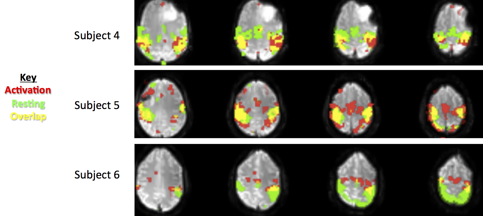
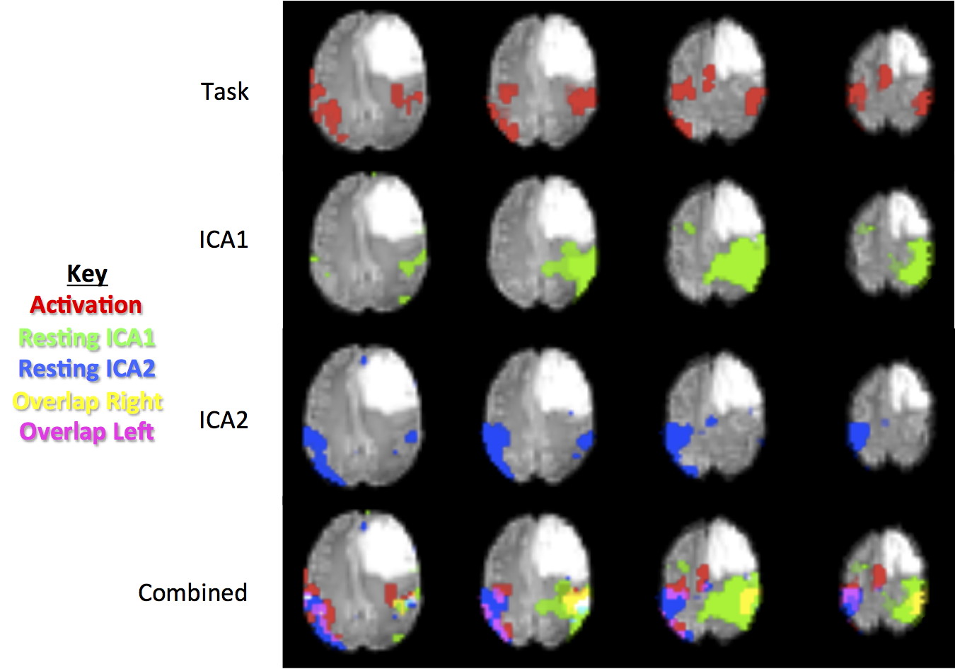
Visual inspection demonstrated resting state maps show good definition of the motor network, in agreement with the task activation maps (Figure 1). Additionally, the second ICA component in some subjects added additional motor network information. In Figure 2, the first ICA component has a larger extent on the side of the pathology, while the second ICA component adds in the contralateral side.
The calculated motor task activation overlap with the resting state network was high across all subjects (0.73±0.14), but the Dice coefficient was low (0.29±0.14), indicating the resting-state networks contain the majority of task activation, but also extend farther spatially.
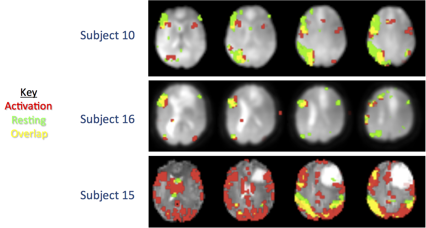
The same analysis was done for the language networks, Figure 3 shows typical results. The resting-state language ICA network has a high spatial overlap with the verb task activation (0.83±0.17).
Conclusions
In this study, we investigated using resting-state fMRI to identify coherent brain networks using a data-driven analysis in presurgical tumor patients. Both motor and language networks were identified from a single resting-state scan, and the resulting network maps compared well to corresponding task activation maps, with a high percent of spatial overlap, without requiring an external task.
These results demonstrate the utility of data-driven resting-state fMRI in presurgical planning of brain tumors as a complement and extension to standard motor and language task activation. Further work will investigate map threshold effects, automatic combination of related ICA components, and investigate the relation to patient outcomes.
Acknowledgements
No acknowledgement found.References
1) Peltier S, Shah Y. 'Biophysical modulations of functional connectivity', Brain Connectivity, 2011, 1:267.
2) Schneider FC, Pailler M, Faillenot I, et al. 'Presurgical assessment of the sensorimotor cortex using resting-state fMRI', AJNR Am J Neuroradiol, 2016, 37:101.
3) Hou B, Bhatia S, Carpenter JS. 'Quantitative comparisons on hand motor functional areas determined by resting state and task BOLD fMRI and anatomical MRI for pre-surgical planning of patients with brain tumors', NeuroImage: Clinical, 2016, 11:378.
4) https://research.ics.aalto.fi/ica/fastica/
5) Cordes D, V.M. Haughton, K. Arfanakis, et al. 'Frequencies contributing to functional connectivity in the cerebral cortex in “resting-state” data', Am. J. Neuroradiol., 2001, 22:1326.
Figures