2080
Integration and segregation of functional segmented anterior and posterior hippocampal networks in memory performance1Radiology, the Second Affiliated Hospital of Zhejiang University, School of Medicine, Hangzhou, China
Synopsis
In this study, we used a novel functional segmentation method to subdivided the left and right hippocampus into anterior and posterior portions according to preferred functional connections with certain cortical regions. And we investigated the association between specific performance of verbal and visual memories and intra-hemispheric resting state FC across anterior and posterior hippocampal networks using resting functional MRI measures in healthy young volunteers. The present results demonstrated that, the anterior hippocampus was specifically involved in the visual memory processing, whereas the posterior hippocampus contributed to both the verbal and visual memories, which may have implications for a functionally synergetic and dissociable role of the hippocampus in different kinds of memory.
Purpose
The interaction between the hippocampus and cortical regions is critical for memory functions1-4. Accumulating evidence from both animal and human fMRI research indicated that the anterior and posterior portions of the hippocampus formed two functionally distinct memory circuits, respectively connected to different cortical regions, and extended networks that support different forms of memory5, 6. Although studies have revealed different memory association along the anterior-posterior axis of the hippocampus, it is unclear how the anterior and posterior hippocampal networks participate and work in division and cooperation in different memory. Moreover, most functional MRI studies have examined the hippocampus subfields with anatomical segmentation. The purpose of the present study was to determine the association between functional connectivity (FC) of functional-segmented anterior and posterior portions of the hippocampus and performance on verbal and visual memory tests in a young, healthy population.Methods
We recruited 100 healthy participants in the age of 19-29 (Mean age=24.03±1.86 years, Male/Female=41/59 education=17.36±1.54 years). Resting state fMRI data were acquired and voxel-wise correlation analysis was performed to functionally divide the hippocampus. We investigated the inter-hemispheric hippocampal-cortical functional connectivity after the participants took the assessment of episodic memory using verbal (California Verbal Learning Test II, CVLT-II) and visual subtests (Rey-Osterrieth Complex Figure, ROCF). The drawing was scored according with the Boston Qualitative Scoring System (BQSS). After preprocessing, fMRI data was further analyzed within the group. The masks of the cortical regions and hippocampus were obtained from the Harvard-Oxford Structural Atlas and Juelich probabilistic standard-brain atlases. We averaged the time-courses for all the voxels in the anterior and posterior hippocampal segment in all participants, respectively, to acquire two seeds of each hemisphere as referenced anterior and posterior hippocampal time-course. The partial correlation coefficient was calculated between the seed reference and its associated hemispheric cortical region regressing out the other cortical time series in the associated hemisphere. Paired t-tests were used to compare the asymmetry of the hippocampal mean correlation values between bilateral anterior and posterior hippocampal networks. The partial correlations were used to identify the association between the intra-hemispheric hippocampal-cortical mean resting correlation and memory performance (verbal and visual memory) by controlling covariance (age, gender and education).Results
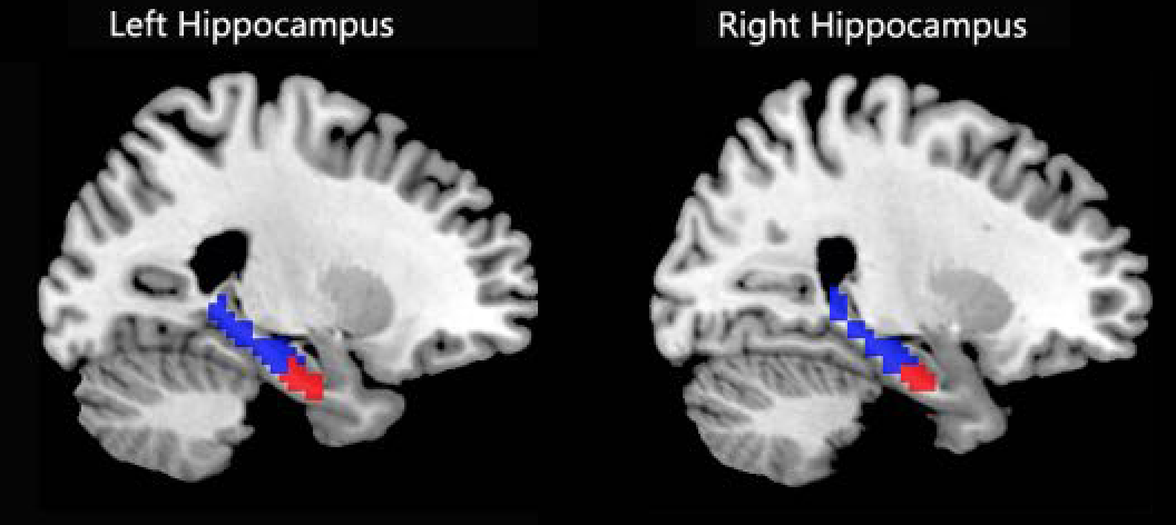
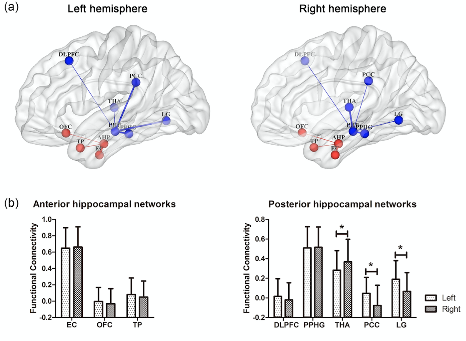
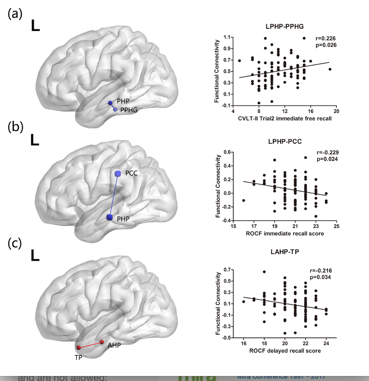
The functional segmentation of bilateral hippocampus demonstrated a large posterior portion and a small anterior portion along the long axis of the hippocampus (Fig. 1). In both hemispheres, the EC and PPHG showed the strongest connection with anterior and posterior hippocampus, respectively (Fig. 2). We found that the resting FC between posterior hippocampus and LG, PCC in the left hemisphere were stronger than those in the right hemisphere (paired t-test, p<0.001). In addition, the resting FC between posterior hippocampus and THA in the right hemisphere was stronger than that in the left hemisphere (paired t-test, p<0.001). We tested whether altered resting FC between anterior and posterior hippocampus and accordingly cortical regions was functionally reflected in performance during verbal and visual memory after correcting for age, gender and education. The results showed that the anterior and posterior hippocampal networks involved differently in verbal and visual memory (Fig. 4). Intra-hemispheric FC between left posterior hippocampus and posterior parahippocampal gyrus (PPHG) was positively correlated with CVLT-II Trail 2 Immediate Free Recall (r=0.226, p=0.026). Intra-hemispheric FC between left posterior hippocampus and posterior cingulate (PCC) was negatively correlated with ROCF Immediate Recall (r=-0.229 p=0.024). Intra-hemispheric FC between left anterior hippocampus and temporal pole (TP) negatively correlated with ROCF Delayed Recall (r=-0.216, p=0.034).Discussion and Conclusion
Based on the different
memory association along the anterior-posterior axis of the hippocampus, we raised
a hypothesis that the functions of anterior and posterior hippocampal networks
in verbal and visual memory were integrated and segregated.
The present results demonstrated that,
only posterior hippocampal networks correlated
with verbal memory, while both anterior and posterior hippocampal networks
involved in visual memory; posterior hippocampal networks appeared more
important in immediate recall, yet the anterior hippocampal
networks were associated with remembering long-delayed visual contents, which
may have implications for a functionally synergetic and dissociable role of the
hippocampus in different kinds of memory.
Previous research suggested
functional difference between anterior and
posterior hippocampus in memory5.
However,
hippocampal subfields are intricately interconnected7.
Our connectivity analysis results providing the
first evidence for functional integration in hippocampal memory networks and
further support for the functional dissociation between anterior and posterior hippocampus.Acknowledgements
Thanks to my colleague Zhujing Shen and my husband for supporting my research work.References
1. Teyler TJ, DiScenna P. The role of hippocampus in memory: a hypothesis. Neurosci Biobehav Rev. 1985 Fall;9(3):377-389.
2. Squire LR, Alvarez P. Retrograde amnesia and memory consolidation: a neurobiological perspective. Curr Opin Neurobiol. 1995 Apr;5(2):169-177.
3. Nadel L, Moscovitch M. Memory consolidation, retrograde amnesia and the hippocampal complex. Curr Opin Neurobiol. 1997 Apr;7(2):217-227.
4. Dash PK, Hebert AE, Runyan JD. A unified theory for systems and cellular memory consolidation. Brain Res Brain Res Rev. 2004 Apr;45(1):30-37.
5. Ranganath C, Ritchey M. Two cortical systems for memory-guided behavior. Nat Rev Neurosci. 2012 Oct;13(10):713-726.
6. Voets NL, Zamboni G, Stoke MG, Carpenter K, Stacey R, Adcock JE. Aberrant functional connectivity in dissociable hippocampal networks is associated with deficits in memory. J Neurosci 2014; 34:4920-4928.
7. Lavenex P, Banta Lavenex P. Building hippocampal circuits to learn and remember: insights into the development of human memory. Behav Brain Res. 2013 Oct 1;254:8-21.
Figures