2079
Development of Individual Evaluation System for White Matter Hyperintesity Recognition Using Deep Convolutional Neural NetworkKyung Mi Lee1, Hyug-Gi Kim2, Jiwon Yoon2, Mi-hyun Kim3, Jang-Hoon Oh3, In Young Lee3, Soonchan Park4, Chang-Woo Ryu4, Eui Jong Kim1, Woo Suk Choi1, Na Rae Yang5, and Jihye Song 6
1Kyung Hee University College of Medicine, Kyung Hee University Hospital, Seoul, Republic of Korea, 2Kyung Hee University Hospital, Seoul, Republic of Korea, 3Univeristy Industry Cooperation, Kyung Hee University, Seoul, Republic of Korea, 4Kyung Hee University Hospital at Gangdong, Seoul, Republic of Korea, 5Neurosurgery, Ewha Womans University School of Medicine, Mokdong Hospital, Seoul, Republic of Korea, 6College of Medicine, Konyang University Hospital, Konyang University Myunggok Medical Research Institute, Daejeon, Republic of Korea
Synopsis
White matter hyperintensity (WMH) is one of the important characteristics of cerebral small vessel disease (cSVD). The objective of this study to investigate the feasibility of WMH recognition using deep convolutional neural networks (CNN). Furthermore, individual evaluation system was proposed to classify WMH groups.
Synopsis
White matter hyperintensity (WMH) is one of the important characteristics of cerebral small vessel disease (cSVD). The objective of this study to investigate the feasibility of WMH recognition using deep convolutional neural networks (CNN). Furthermore, individual evaluation system was proposed to classify WMH groups.Introduction
The white matter hyperintensities (WMH) are abnormal increased signal intensities seen in white matter regions within the cerebrum and brainstem on FLAIR images1. WMH has a crucial role in many neurologic fields including stroke, dementia and normal aging process2. However it is difficult to representative the characteristic of WHM degree automatically because WMH in the older population may be small, diffuse and irregular in shape and sufficiently heterogeneous across subjects. According to the previous studies, WMHs have potential prognostic value for brain aging evaluation, but they are not routinely employed as a diagnostic measure in clinical practice. Performing a comprehensive manual counting of number and distribution of lesions in the clinical setting is simply not practical. And another reason is in previous studies account the calculation system for the multiple sclerosis application, WMH extraction methodology is different from the normal aging population. The objective of this study to investigate the feasibility of WMH recognition using deep convolutional neural networks (CNN). This work presents an automated framework for quantification of WMHs in multi-modal MRI using the deep learning technique.Materials and Methods
1,511 elderly healthy subjects (mean age = 70.66) were participated after informed consent. All subjects were scanned on the 3T MR (Achieva, Philips Healthcare, Best, Netherlands) with 2D-axial FLAIR sequences. The WMH score is a 4-point scale (none, mild, moderate and severe) 3. Furthermore, all subjects’ WMH scores using logistic regression with a dichotomized score: non-advanced group (none or mild WMH) and advanced group (moderate and severe WMH). All pre-processing was performed in Matlab software (MathWorks, Natick, MA, USA). Input dataset was pre-processed for data argumentation. Pre-processing of the training data-set was evaluated as follows: size normalization of input data as 227x227 matrix and for data argumentation, image was rotated 4 direction (0, 90, 180, and 270 degree, fig 1). We implemented on the data to differentiate among WMH groups with the AlexNet CNN model (5 convolution layers and 3 fully-connected layers, fig 2) that is a powerful deep learning architecture for imaging classification4. All processing was performed with 8 GB GPU environment. The result of classification were quantitatively assessed by accuracy and evaluated using the activated feature maps in each layer. Two radiologists were reviewed all classified images to validate labeling.Results
WMH groups were classified from result the review of two neuroradiologists (kappa= 0.8). The accuracy of the set of training data with deep CNN model was 99.5% and the result of classification with the set of test data showed that the accuracy was 77.3% for four labeled groups. The highest accuracy was noted in the severe groups and the lowest accuracy was presented in the mild degree group. Performance evaluation was done by using the confusion matrix and ROC curve analysis (fig 3). Learned WMH classifier by deep learning recognized high intensity as feature of WMH. These activated features of the WMH were visually confirmed to be closely correlated with the features evaluated FLAIR MR image reading by radiologists.Discussions
Deep learning technique has advanced rapidly in recent years by growing of big data process. Especially, the medical imaging field is essential role because it provides useful information. One of the main advantage is that individual diagnosis is possible for specific disease. Present WMH diagnosis was evaluated by a radiologist’s experience. For more objective and quantitative estimation, in this study, we proposed the novel classification method using deep learning algorithm for WMH evaluation. The highest accuracy was noted in the severe degree group, probable due to easy performance. This means that the burden of WMH is relative appropriate and number of lesions are small. In other words, in the mid degree group, the accuracy is low because the high signal intensity spot is difficult to be detected and extract volume is not clarify to cut off between the mild and the moderate degree. We have some limitations. First, we will be obtain the more learning data. Second, we need to optimize deep learning model. For these, we modified of ‘AlexNet’ model to reduce the overfitting and develop of novel deep learning model for generalized model without pre-processing step. Last, we need more information from deep learning algorithm. We called this ‘white-box project’ - to descript the result of deep learning using ‘black-box’ opening.Conclusion
The deep CNN based on personalized WMH evaluation system proved to be more effective the degree of WMH process.Acknowledgements
This work was supported by Basic Science Research Program through the Ministry of Education of the Republic of Korea (NRF-2016R1D1A1B03933173) and by the National Research Foundation of Korea (NRF) grant funded by the Korea Government (MSIP) (NRF-2017R1E1A2A02067113).References
1. Thompson CS et al. Stroke 2009;40:e322-30. 2. Staals J et al. Neurobiol Aging 2015;36:2806-11. 3. Inzitari et al. BMJ. 2009;6:339. 4. Alex Krizhevsky et al. http://code.google.com/p/cuda-convnet/.Figures
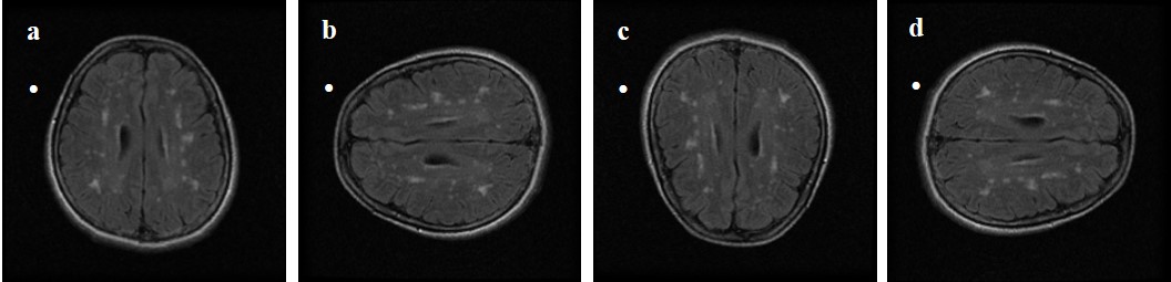
Fig 1. Image argumentation using
rotation. (a) original image – 0 degree, (b) 90 degree, (c) 180 degree, (d) 270
degree rotated images.
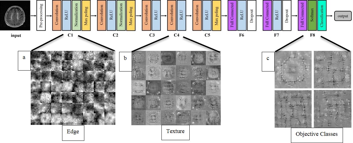
Fig 2. AlexNet CNN arcitecture and feature
maps in each layer. Major feature map in (a) 1st convolution layer,
(b) 4th convolution later and (c) final fully connected layer.
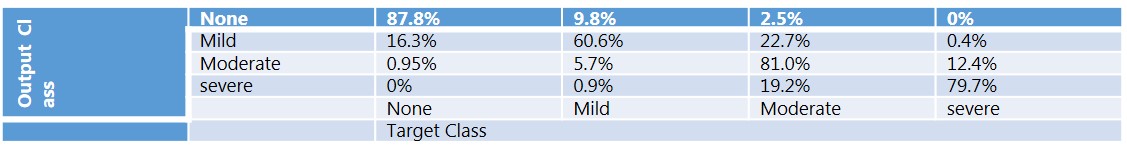
Fig 3. Performance evaluation.
Evaluation of accuracy using confusion matrix.