2078
Altered Intrinsic Brain Activity and Memory Function Improvement in Patients with End-Stage Renal Disease During A Single Dialysis SessionPeng Li1, Dun Ding2, Xueying Ma2, Huawen Zhang1, Jixin Liu3, and Ming Zhang2
1Department of Medical Imaging, NO.215 Hospital of Shaanxi Nuclear Industry, Xianyang, China, 2Department of Medical Imaging, First Affiliated Hospital of Xi’an Jiaotong University, Xi’an, China, 3Center for Brain Imaging, School of Life Science and Technology, Xidian University, Xi'an, China
Synopsis
The underlying neural mechanisms of the memory deficits in end-stage renal disease patients with dialysis treatment are poorly understood. Here we analyzed the resting-state brain activity changes and the related memory improvement by using mALFF and ReHo methods before dialysis (T1pre-dialysis) and after 24 hours (T2post-dialysis). The results indicated that regional spontaneous activity changes of the DLPFC were related with memory improvement after a single dialysis treatment, which may provide insight into the effect of hemodialysis on changes of brain function and cognitive impairments.
Introduction
End-stage renal disease (ESRD) is defined as the estimated glomerular filtration rate less than 15 mL/min/1.73m2 demanding maintenance dialysis therapy or kidney transplantation.1 Memory deficits were considered to have a great influence on self-management, dietary restriction and therapeutic regimen for ESRD patients with dialysis treatment.2, 3 Most of previous studies only focused on the neuropsychological scales, lacking hard evidence to determine the potential neuromechanism influence of dialysis treatment on memory deficits.4 The aim of our study is to investigate changed spontaneous brain activity and its relationship with altered memory performance in patients with end-stage renal disease before dialysis and after 24 hours during a single dialysis session.Methods
By using resting functional magnetic resonance imaging (rs-fMRI), 23 ESRD patients (age: 34 ± 8.7 years, dialysis age: 31.3 ± 20.7 month) and 25 matched healthy controls (HCs) (age: 33 ± 10.3 years) were scanned before dialysis at baseline (T1pre-dialysis), and all patients were also scanned after 24 hours (T2post-dialysis) in a single dialysis session. Brain scanning was obtained on a GE 3.0-T MR scanner (Discovery MR 750) with an 8-channel phase array head coil. Individual rs-fMRI data was collected using echo-planar imaging (EPI) sequence. High-resolution anatomical image was collected using a 3D Brain Volume imaging sequence (BRAVO). Amplitude of low-frequency fluctuation (ALFF) and regional homogeneity (ReHo) methods were used to evaluate the between-group differences. The Auditory Verbal Learning Test–Huashan version (AVLT-H) was performed to assess an individual’s memory function. For statistical analysis, the differences of the mean ALFF (mALFF) values and ReHo were tested using a two-sample t-test between ESRD patients and HCs at baseline after controlling age and gender. The difference in mALFF values and ReHo between T1pre-dialysis and T2post-dialysis were tested using a paired-sample t-test. The baseline memory function performances differences were evaluated using a two-sample t-test and the differences between T1pre-dialysis and T2post-dialysis were compared with a paired-sample t-test. Age and education level were regressed as covariates to remove their impact. After that, correlations between changed rs-fMRI data and altered AVLT-H subscale score were conducted were assessed by Pearson correlation analyses.Results
At baseline compared with the HCs, ESRD patients showed decreased mALFF values mainly located in the dorsolateral prefrontal cortex (DLPFC), medial frontal gyrus, and precuneus (Fig.1). The significant ReHo increased in the bilateral inferior temporal gyrus and left hippocampus, but decreased in the right precentral gyrus and anterior cingulate cortex (Fig.2). At T2post-dialysis compared with T1pre-dialysis, the mALFF values of the DLPFC and precuneus were increased (Fig.3.a). No significantly different ReHo region was found during a dialysis session. Memory function performances showed a significant decrease in the immediate recall total score (IR-S), long-term delayed recall score (LR-S), and recognition score (REC-S) at baseline. The IR-S, LR-S, and REC-S were significantly increased at T2post-dialysis. Additionally, the increased mALFF values of the DLPFC were significantly positively correlated with the improvement of IR-S (Fig.3.b).Discussion
By using rs-fMRI, our results showed that increased spontaneous brain activity regions were mainly in the DLPFC and precuneus at 24 hours after dialysis compared with pre-dialysis patients. A positive correlation between the increased mALFF values in the DLPFC and the increased IR-S was observed. Our findings showed that dialysis treatment could modulate intrinsic brain activity and improve memory function after a dialysis session. The regional spontaneous activity changes of the DLPFC may reflect verbal working memory improvement at 24 hours after dialysis in ESRD patients.5,6 We indicated the intrinsic activity changes of the precuneus and DLPFC by relative removal of the neurotoxin after a single dialysis treatment , which may provide insight into the effect of hemodialysis on spontaneous brain function during a single dialysis session.7,8Conclusion
Our results indicated that regional spontaneous activity changes of the DLPFC might reflect memory performance improvement after a single dialysis treatment, which may provide more information to reveal the effect of hemodialysis on changes of brain function and cognitive impairments.Acknowledgements
This study was funded by the National Natural Science Foundation ofChina (Grant No. 81371530) and the Research Funds of the First Affiliated Hospital of Xi’anJiaotong University, College of Medicine (Grant No. 2014YK3).References
- Bugnicourt J M, Godefroy O, Chillon J M, et al. Cognitive disorders and dementia in CKD: the neglected kidney-brain axis. J Am Soc Nephrol, 2013. 24(3): 353-63.
- Campbell N L, Boustani M A, Skopelja E N, et al. Medication Adherence in Older Adults With Cognitive Impairment: A Systematic Evidence-Based Review. Am J Geriatr Pharmacother, 2012. 10(3): 165-77.
- Elias M F, Dore G A, Davey A. Kidney disease and cognitive function, Kidney disease and cognitive function. Contrib Nephrol, 2013. 179: 42-57.
- Schneider S M, Malecki A K, Müller K, et al. Effect of a single dialysis session on cognitive function in CKD5D patients: a prospective clinical study. Nephrol Dial Transplant, 2015. 30(9): 1551-9.
- Fried P J, Rd R R, Moss M B, et al. Causal Evidence Supporting Functional Dissociation of Verbal and Spatial Working Memory in the Human Dorsolateral Prefrontal Cortex. European Journal of Neuroscience, 2014. 39(11): 1973-81.
- Wolk D A, Dickerson B C. Fractionating verbal episodic memory in Alzheimer's disease. Neuroimage, 2011. 54(2): 1530-9.
- Ni L, Wen J, Zhang L J, et al. Aberrant default-mode functional connectivity in patients with end-stage renal disease: a resting-state functional MR imaging study. Radiology, 2014. 271(2): 543-52.
- Zhang L J, Wen J, Liang X, et al. Brain Default Mode Network Changes after Renal Transplantation: A Diffusion-Tensor Imaging and Resting-State Functional MR Imaging Study. Radiology, 2015, 278(2): 485.
Figures
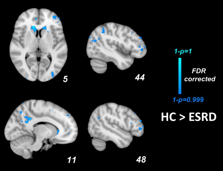
Figure1. Mean ALFF maps show differences among patients at T1pre-dialysis and healthy control subject (HCs)
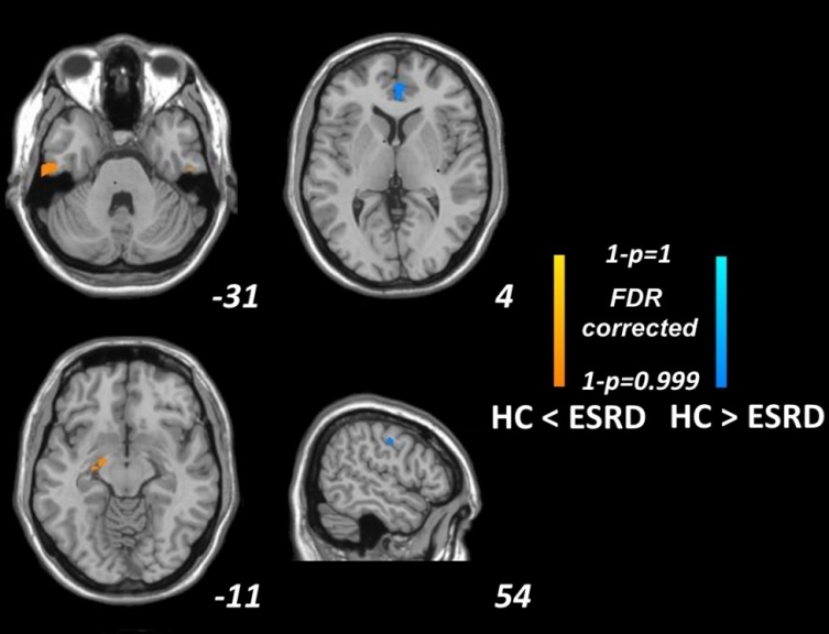
Figure 2. ReHo differences among patients at T1pre-dialysis and healthy control subject (HCs)
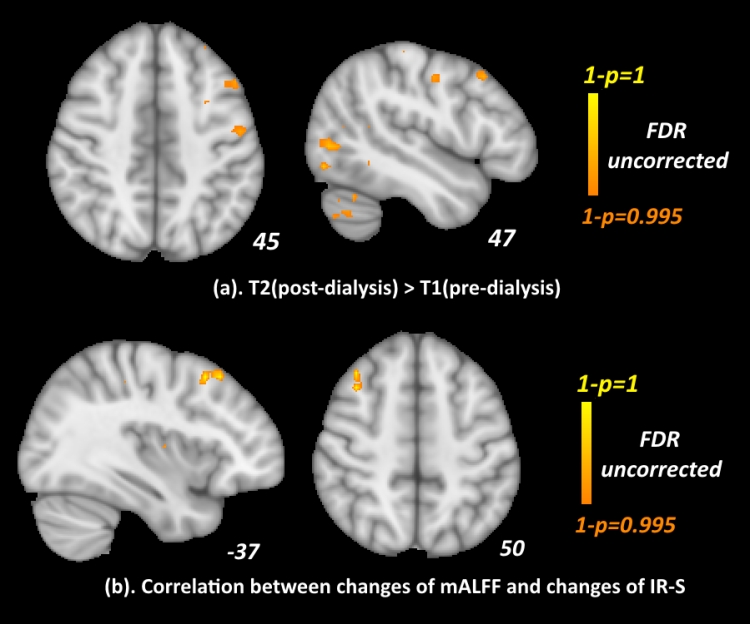
Figure3. (a) Mean ALFF maps show increased mean ALFF regions in patients at T2post-dialysis relative to T1pre-dialysis (b) Mean ALFF maps show correlation between changes of mean ALFF values region and changes of IR-S in patients at T2post-dialysis relative to T1pre-dialysis.