2077
Vessel Wall Thickness Evolution Across the Lifespan Assessed using Non-invasive Intracranial Vessel Wall Imaging1Vanderbilt University Medical Center, Nashville, TN, United States
Synopsis
Vessel wall imaging is becoming more widely applied, however normal, age-specific ranges for wall thickness have not been established. We applied a variable refocusing angle 3D-TSE acquisition with and without a DANTE flow suppression module to healthy subjects (ages=8-79 years; n=82). Vessel wall measurements revealed no significant change in wall thickness with age for the supraclinoid internal carotid arteries and basilar artery. The outer wall diameter and wall thickness were measured to be less for the acquisition with versus without DANTE. These data suggest that unlike tissue volume, vessel wall thickness is relatively constant across the lifespan for healthy subjects.
Introduction
Intracranial vessel wall imaging (VWI) has recently become available and may be used to assess a variety of intracranial vascular diseases (1). Detection of vessel wall disease with VWI may include evaluation of wall thickness among other characteristics such as wall morphology, signal and enhancement. To detect abnormal wall thickening in patients with cerebrovascular disease that manifests at different ages, e.g., sickle cell anemia (5-50 years) (2), moyamoya (5-50 years) (3), or atherosclerotic arterial steno-occlusion (> 45 years) (4), it is necessary to have a reference value, especially in diseases that lead to circumferential wall thickening. Prior studies evaluating wall thickness in healthy subjects have included a broad range of ages (5), and while tissue atrophy (6) with age has been documented, it remains uncertain how wall thickness changes with age. Additionally, it is unclear how wall thickness measurements vary with the use of different vessel wall imaging techniques, such as different blood or CSF suppression methods. The purpose of this study is to evaluate vessel wall thickness in a cohort of healthy subjects free from cerebrovascular disease to (i) better establish reference wall thicknesses across the lifespan, (ii) determine if vessel wall thickness changes across the lifespan for healthy aging, and (iii) determine if wall thickness measurements vary with vs. without novel flow suppression modules (7,8).Methods
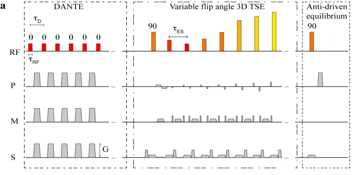
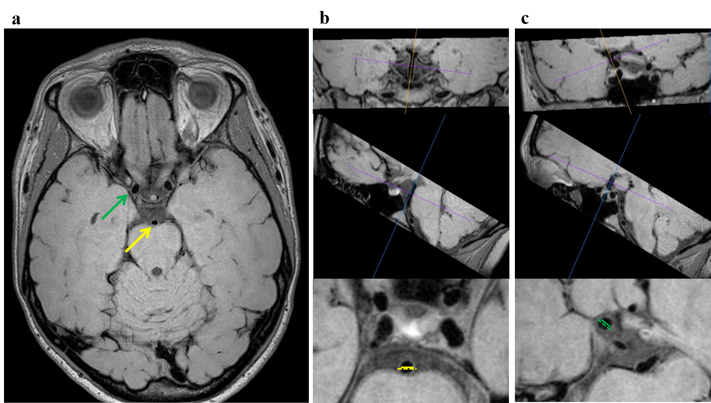
Subjects: Healthy subjects (n=82; gender= 42/40 male/female; age range=8-79 years) provided informed, written consent for this HIPAA-compliant, IRB-approved study. Approximately 10 subjects were imaged per decade of life. Imaging experiments: Scans were performed at 3.0T (Philips) using body coil transmission and a 32-channel receive head coil. Two vessel wall acquisitions were performed: (i) a proton density-weighted 3D-TSE acquisition (Figure 1) with axial orientation, variable refocusing angle pulse sweep=40-120°, TSE factor=56, TR/TE=1500/33ms, field-of-view(FOV)=200x166x45mm3, spatial resolution=0.5x0.5x1.0mm3, SENSE factor=2.0, and duration=4min42s and (ii) the same acquisition with the addition of the DANTE module (7) for improved CSF suppression (DANTE parameters repetitions=300, interval=1.1ms, gradient strength in three directions=22.5mT/m, flip angle=8˚). Analysis: Vessel wall measurements were performed by two independent radiologist readers blinded to subject age using Osirix (Pixmeo). In both vessel wall scans, the luminal diameter and outer vessel wall diameter were measured in the supraclinoid internal carotid arteries (ICAs) and the basilar artery. Measurements were made in the left-right direction in a plane perpendicular to the vessel length using Osirix multi-planar reformatting tool (Figure 2). The wall thickness was calculated as one half the difference of the outer vessel wall and luminal diameters. Statistical considerations: Linear regression was performed to evaluate the relationship between vessel wall thickness and age, in each of the measured vessel segments, with and without DANTE. A paired t-test was used to evaluate differences in wall measurements between the acquisitions without and with DANTE for each vessel segment among all subjects.Results
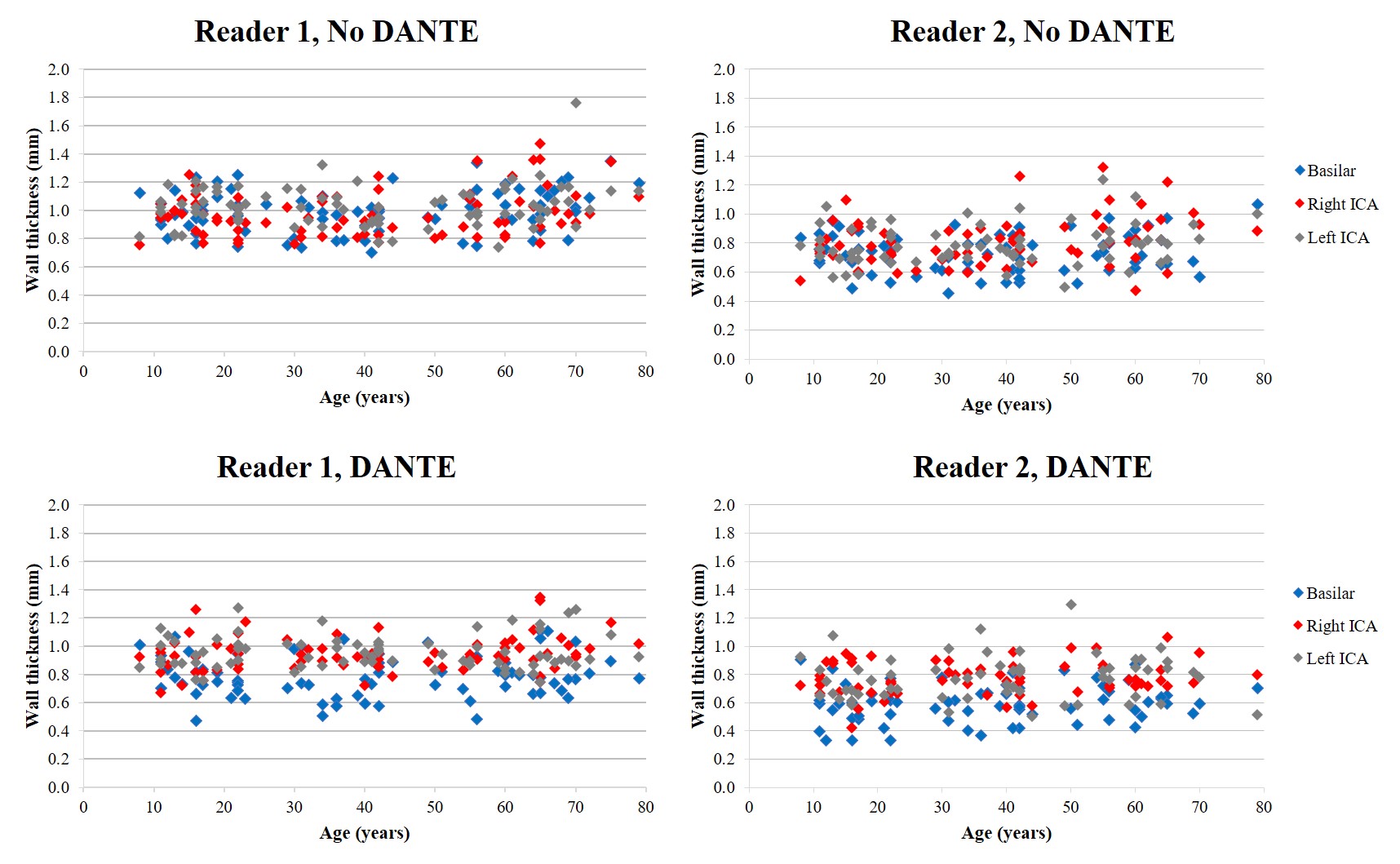
Figures 3 shows wall thickness versus age for each measured vessel wall segment, with and without DANTE, separately for the two readers. Although absolute wall thickness values differed slightly between readers, linear regression showed no significant change in wall thickness with age for either reader. For both readers the wall thickness measurement was less (p<0.05) with vs without DANTE in the basilar artery (reader 1: 0.80±0.17mm versus 1.04±0.16mm, reader 2: 0.59±0.13mm vs 0.73±0.14mm) and showed a trend toward decreased wall thickness with DANTE in the right and left ICAs. Wall thickness differences between acquisitions were attributed primarily to a decrease in the outer vessel wall diameter measurement with DANTE. Finally, correlation between readers was significantly (p<0.05) improved with DANTE versus without and for the luminal diameter versus the outer wall diameter measurement.Discussion
We assessed vessel wall measurements in healthy subjects across the lifespan and found no difference in wall thickness with age at the spatial resolution available with current intracranial VWI approaches. Addition of the DANTE CSF suppression module showed on average a decrease in the outer wall diameter and wall thickness. The difference in measurements was greatest for the basilar artery, which may be due to the large size of the CSF cisterns about the basilar artery and greater effect of CSF suppression in that region. In some cases, DANTE may also cause loss of signal in the vessel wall, artifactually decreasing its apparent thickness.Conclusion
Vessel wall imaging without and with CSF suppression modules shows no difference in wall thickness with age in a group of healthy subjects spanning eight decades of life. The outer wall diameter and wall thickness are on average less with the use of CSF suppression modules.Acknowledgements
No acknowledgement found.References
1. Mandell DM, Mossa-Basha M, Qiao Y, et al. Intracranial Vessel Wall MRI: Principles and Expert Consensus Recommendations of the American Society of Neuroradiology. Am. J. Neuroradiol. [Internet] 2016. doi: 10.3174/ajnr.A4893.
2. Ohene-Frempong K, Weiner SJ, Sleeper LA, Miller ST, Embury S, Moohr JW, Wethers DL, Pegelow CH, Gill FM, Disease the CS of SC. Cerebrovascular Accidents in Sickle Cell Disease: Rates and Risk Factors. Blood 1998;91:288–294.
3. Scott RM, Smith ER. Moyamoya Disease and Moyamoya Syndrome. N. Engl. J. Med. 2009;360:1226–1237. doi: 10.1056/NEJMra0804622.
4. Jacobs BS, Boden-Albala B, Lin I-F, Sacco RL. Stroke in the young in the northern Manhattan stroke study. Stroke 2002;33:2789–2793.
5. Qiao Y, Steinman DA, Qin Q, Etesami M, Schär M, Astor BC, Wasserman BA. Intracranial arterial wall imaging using three-dimensional high isotropic resolution black blood MRI at 3.0 Tesla. J. Magn. Reson. Imaging 2011;34:22–30. doi: 10.1002/jmri.22592.
6. Peters R. Ageing and the brain. Postgrad. Med. J. 2006;82:84–88. doi: 10.1136/pgmj.2005.036665.
7. Li L, Miller KL, Jezzard P. DANTE-prepared pulse trains: a novel approach to motion-sensitized and motion-suppressed quantitative magnetic resonance imaging. Magn. Reson. Med. 2012;68:1423–1438. doi: 10.1002/mrm.24142.
8. Wang J, Helle M, Zhou Z, Börnert P, Hatsukami TS, Yuan C. Joint blood and cerebrospinal fluid suppression for intracranial vessel wall MRI. Magn. Reson. Med. 2016;75:831–838. doi: 10.1002/mrm.25667.
Figures