2055
Whole Tumor Histogram Analysis of T2-Weighted, Diffusion-weighted, and Postcontrast T1-Weighted Images in Medulloblatoma:Assessment Risk of Recurrence.1the First Affiliated Hospital of Zhengzhou University, zhengzhou, China
Synopsis
Retrospective analysis of 28 patients which were pathologically confirmed medulloblastoma.We find that MRI whole-tumor histogram analysis can be used as an important supplementary method to assess the risk of medulloblastoma recurrence.
Objective
To explore the value of MRI whole-tumor histogram in the risk assessment of medulloblastoma recurrence.Methods
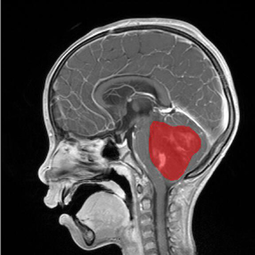
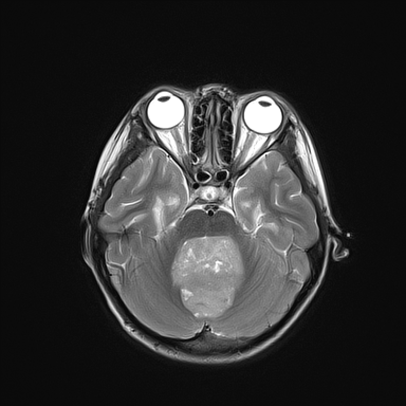
Retrospective analysis of 28 patients which were pathologically confirmed medulloblastoma.These patients were divided into two groups,which were defined as the recurrent group and the non recurrent group.Using the software Mazda,whole-tumor regions of interest were drawn on all slices of diffusion-weighted, T2-Weighted, and Postcontrast T1-Weighted Images to obtain histogram parameters,including the average value, variance, skewness, kurtosis, perc.01%,perc.10%,perc.50%,perc.90%,perc.99%.The histogram parameters were analyzed statistically to find out the characteristics of the significant differences between the two groups. Next, drew the ROC curve to assess diagnostic efficiency in medulloblastoma recurrenceResults
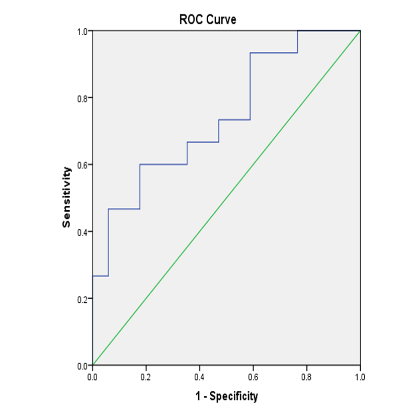
The variance, the kurtosis , the 90th and 99th percentiles derived from postcontrast T1-weighted images have the statistical significance (P<0.05).The variance showed the sensitivity and the specificity of the risk of medulloblastoma recurrence were 84.6% and 61.5% respectively; as for kurtosis,there were 61.5% and 76.9%, the 90th percentile suggested both the sensitivity and the specificity were 69.2%; and by the 99th percentile, they were 69.2% and 61.5% respectively;The variance derived from T2-Weighted images have the statistical significance (P<0.05).the sensitivity and the specificity were 66.7% and 64.7%; However,there is no difference of parameters between two groups in the ADC (P>0.5).Conclusion
MRI whole-tumor histogram analysis can be used as an important supplementary method to assess the risk of medulloblastoma recurrence.The variance derived from precontrast T1-weighted images has the potential to be the most significant parameter for assessmenting the risk of medulloblastoma recurrence.Acknowledgements
no
References
[1]Xu XQ,Hu H,Su GY,et al.Utility of histogram analysis of ADC maps for differentiating orbital tumors.Diagn Interv Radiol.2016,22(2):161-167
[2]Falk A,FahlstrOm M,Rostrup E,et al.Discrimination between glioma grades II and III in suspected low-grade gliomas using dynamic contrast-enhanced and dynamic susceptibility contrast perfusion MR imaging:a histogram analysis approach.Neuroradiology,2014,56:1031-1038.
[3]Li Z,Mao Y,Huang W,et al.Texture-based classification of different single liver lesion based on SPAIR T2W MRI images.BMC Med Imaging.2017,17(1):42-42
[4]Meyer HJ;Schob S;Münch B,et al.Histogram Analysis of T1-Weighted, T2-Weighted, and Postcontrast T1-Weighted Images in Primary CNS Lymphoma: Correlations with Histopathological Findings-a Preliminary Study.Mol Imaging Biol.2017,DOI:10.1007/s11307-017-1115-5.
Figures