2052
An 8 channel Rhesus Head coil for Neuroimaging on 3TJo Lee1,2, Xing Yang3, Qiaoyan Chen1,2, Changjun Tie1,2, Xiaoliang Zhang4,5, Hairong Zheng1,2, and Ye Li1,2
1Lauterbur Imaging Research Center, Shenzhen Institutes of Advanced Technology, Chinese Academy of Sciences, Shenzhen, China, 2Shenzhen Key Laboratory for MRI, Shenzhen, China, 3High-Field Magnetic Resonance Brain Imaging Key Laboratory of Sichuan Province, School of Life Science and Technology, University of Electronic Science and Technology of China, Chengdu, China, 4Department of Radiology and Biomedical Imaging, University of California San Francisco, San Francisco, CA, United States, 5UCSF/UC Berkeley Joint Graduate Group in Bioengineering, San Francisco, CA, United States
Synopsis
In this study, a custom-designed 8-channel monkey coil was made to match the specific stereotaxic instrument and also better fit the shape of rhesus monkey brain. In comparison with a commercially available coil array, monkey brain images acquired using the dedicated monkey coil array at 3T achieve better SNR, improved parallel imaging capability and higher spatial resolution.
Introduction
Rhesus monkey is a common model used functional neuro MR imaging (fMRI) [1,2]. Due to the required stereotaxic instrument in monkey MR imaging, the commercially available RF coils do not fit the experiment setup. Consequently the imaging signal-to-noise ratio (SNR) and base resolution are dramatically degraded. In this work, we designed, constructed and tested a dedicated eight-channel monkey RF coil array perfectly fitting the MRI stereotaxic instrument and rhesus monkey’s head. The results show that the proposed monkey coil obtained over 66% higher SNR than that of the commercially available Siemens flex small4 in phantom studies, and achieves 0.4×0.4×0.4mm3 high resolution images in in-vivo study at 3T.Methods
The outer appearance of the customized rhesus monkey head coil (monkey coil) was built to perfectly fit the structure of the MRI stereotaxic instrument (KOPF 1430M), as shown in Figure 1(a). From the front view could see that the monkey coil design has two inclined planes at the right and lift side to match the support rod of the inclined plane. This design not only can ensure the monkey coil placed steadily on the instrument. The whole coil shell was made by polycarbonate. The inner view of monkey coil is illustrated in Figure 1(c) and (d). The coil structure was designed in eight coil arrays with single loop size 75×65 mm2 in average. Coil arrays were made by 6mm thick chopper wire, and were arranged in three rows from head to feet direction with following orders: 3,3,2. Each coil array included three parts of elements layout, separated coil array into three equal length. The three-element layout include one active detune board and two tuning capacitances. All coil arrays were tuned at 123.2MHz, and electromagnetic decoupling among the coil elements was achieved by the overlapping method and low input impedance preamplifiers. Phantom study: All on-system studies were scanned by using Siemens 3T Tim Trio system, and all comparison experiments were performed with the 4 channels small flexible coil array (flex small 4) from Siemens. A customized phantom with 432cm2 content volume was made to mimic the adult rhesus monkey head by using photosensitive resin 3D printer, and was filled by (3.75g NISO4×6H2O + 5g NACL)/1000g H2O. The phantom was placed and fixed in the position the rhesus monkey’s head was located in the stereotaxic instrument. Gradient echo (GRE) sequence with following parameters was used for obtaining signals:TR/TE= 300/10ms, slice thickness = 3mm, FOV = 150×150mm2, acquisition matrix = 256×256, flip angle = 60 degree, bandwidth = 130 Hz/pixel, number of average = 1. While the power set to zero was used to acquire the noise. Images in all three orientations were required. MATLAB was used for the post analysis. The method called root sum of squares (sos) from reference[3] was used for calculating SNR. The inverse g-factor map was calculated with acceleration factor set to 2 and the accelerate direction from right to left, anterior to posterior and head to feet for transverse, sagittal and coronal orientation in order. In-vivo study: An adult rhesus monkey (10 years old, weight 11 kg, male) was scanned for the anatomic images, by T1-weighted 3D MPRAGE sequence with following parameters: TR/TE/TI = 2100ms /2.8 ms/ 1100 ms, FOV = 140×140×77.44 mm3, matrix = 320×320×176, isotropic resolution 0.4×0.4×0.4mm3, flip angle = 8 °, bandwidth = 200 Hz/pixel, echo spacing = 8.8ms, number of average = 1.results
The SNR maps and SNR-curve charts were shown in Figure 2. The 1D profiles of SNR along the dash-lines in the images were plotted in the lower inset of Figure 2. The area of the brain was measured from the adult rhesus monkey in the in-vivo study. From the SNR-curves, monkey coil was 66%, 173% and 114% in average better than flex small 4 in transverse, sagittal and coronal orientations. The inverse g-factor maps were represented in Figure 3. The white dash line represents the brain zone, and the calculated g-factor data were written under. The results indicate the parallel imaging capability of the monkey coil is better than flex small 4. The high spatial resolution (0.4×0.4×0.4mm3) of 3D rhesus monkey brain structural images is demonstrated in Figure 4. The images reveal that the proposed monkey coil can achieve higher quality images even in the deep brain area.Discussion/Conclusion
A custom-designed 8-channel monkey coil which well fits the specified stereotaxic instrument in the monkey imaging was constructed and tested for 3T MR imaging. The monkey coil is characterized with high sensitivity, robustness and easy to use, and is able to provide high resolution 3D images of whole rhesus monkey brain, better acceleration ability, and high SNR in all three directions.Acknowledgements
This work was supported in part by NSFC under Grant No. 61571433,81527901,81527901, 81327801and 81627901. Guangdong Province grants 2014A030313691, 2015B020214006 and 2014B030301013. Youth Innovation Promotion Association of CAS No. 2017415, city grants KQJSCX20160301143250 and JCYJ20170413161314734 and a Pengcheng Scholar Award.References
[1] R. Matthew Hutchison, et al., Frontiers in Neuroanatomy 2012, 6(29): 1-19.
[2] Qian Lv, et al., Biol Psychiatry2015, available online.
[3] B. Keil et al., JMR 2013, 229, 75-89.
Figures
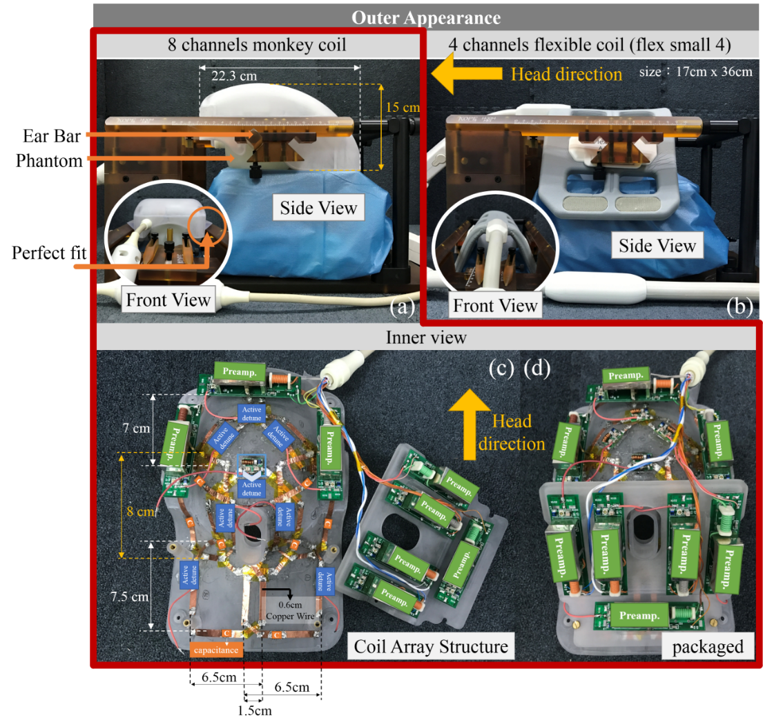
FIgure 1. The photographs and the layouts of
8
channels monkey coil and 4
channels flexible coil (flex small 4).
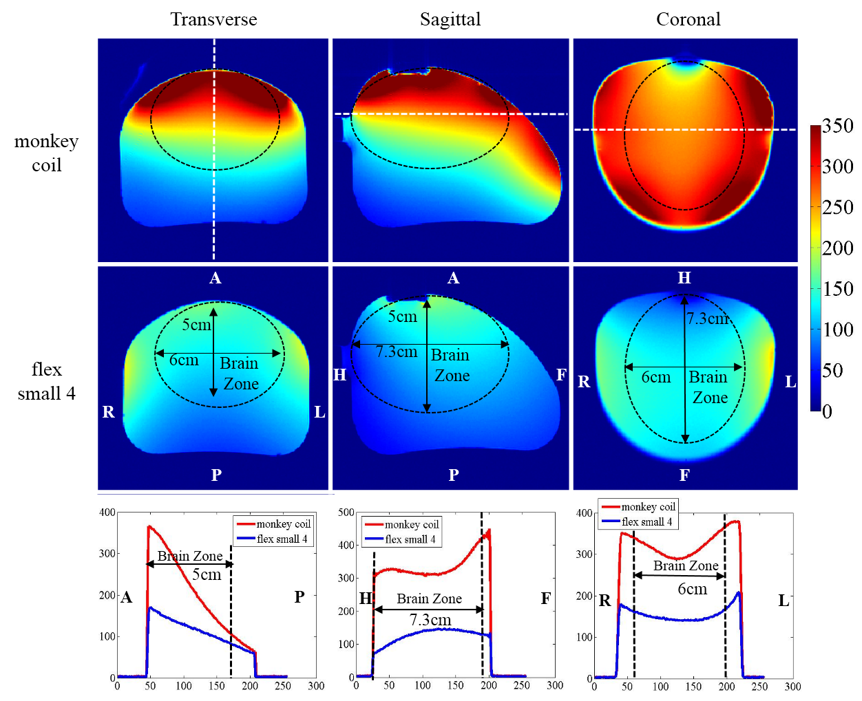
Figure 2. The SNR maps and 1D SNR profiles of the monkey coil
and the flex small 4 in transverse(left column), sagittal(middle column) and coronal(right column) orientations.
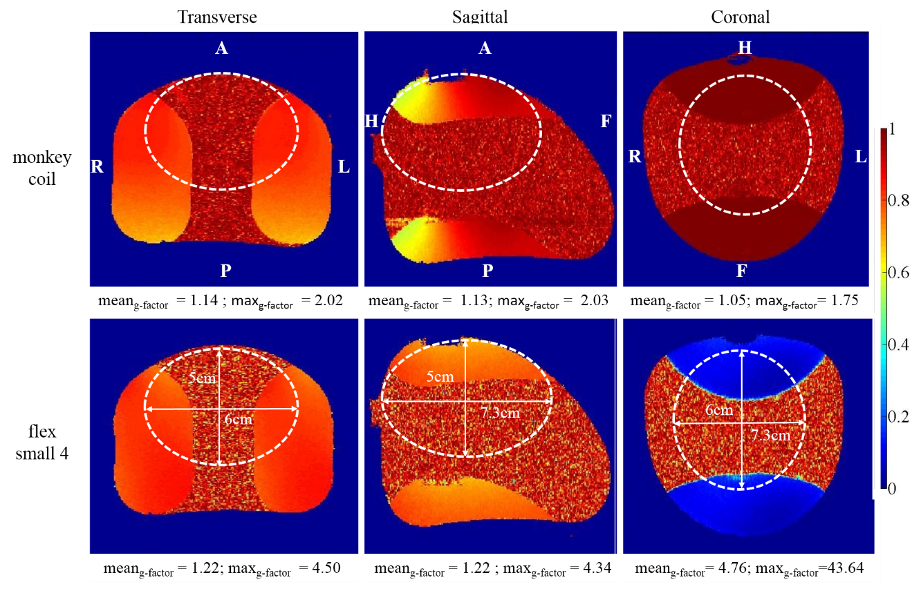
Figure 3. The inverse g-factor maps with acceleration factor = 2 of the monkey coil and the flex
small 4 in three orientations. The g-factor values calculated at the Region of Interest (ROIs) depicted as the while circles were shown at the bottom of the images.
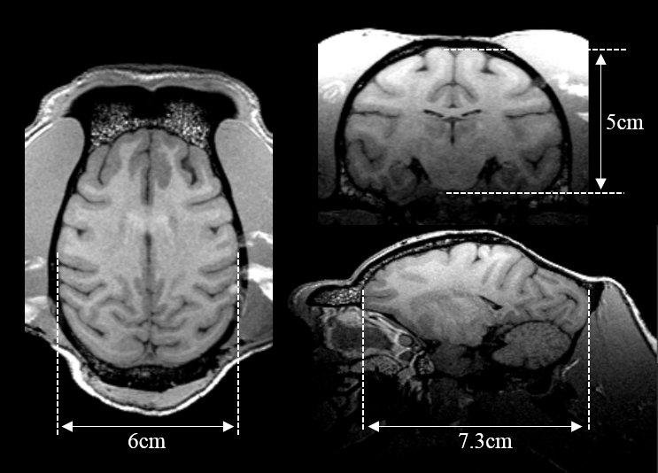
Figure 4. T1 weighted brain images of
the rhesus monkey in the transverse, sagittal and coronal planes with a 0.4×0.4×0.4 mm3 resolution.