2051
Distortion-Free Imaging: A Double Encoding Method (DIADEM), High-Resolution Diffusion Imaging of Brain Tumors on a Compact 3T Scanner1Department of Radiology, Mayo Clinic, Rochester, MN, United States
Synopsis
Recently, we developed a multi-shot method using spin-warp echo-planar encoding technique inspired by point-spread function mapping. Distortion-free imaging: a double encoding method (DIADEM) can achieve distortion-free, very high in-plane spatial resolution whole brain diffusion imaging in less than 10 minutes on a compact 3T scanner with high performance gradients. A clinical feasibility study of brain tumor diffusion imaging was performed to explore the efficacy of this approach compared to standard single-shot, echo-planar imaging commonly used in clinical practice. The results demonstrate that the proposed method allows considerable improvements in characterizing brain tumors especially at regions of the brain typically degraded by high susceptibility artifacts.
Introduction
Diffusion imaging is an important sequence for evaluating brain tumors. However, single-shot echo-planar imaging (ssEPI), which is currently the most commonly used sequence in clinical practice, suffers from limited spatial resolution and high sensitivity to magnetic field inhomogeneity, or susceptibility artifact, that can result in considerable localized geometric distortion. As an alternative, a multi-shot approach1 using hybrid spin-warp and echo-planar encoding strategy has been developed, which allows high-resolution, distortion-free diffusion imaging (DIADEM). Recently, this multi-shot approach has been optimized for a high performance compact 3T imager. DIADEM offers significant scan time reduction and high in-plane spatial resolution using the compact 3T gradient2 and further sequence modification. The purpose of this work was to investigate the clinical feasibility of DIADEM compared to ssEPI in brain tumors.Methods
DIADEM and ssEPI diffusion-weighted images (DWI) were acquired on a compact 3T imager3,4 with a high-performance gradient capable of 80 mT/m sustained amplitude and a slew rate of 700 T/m/s using either an 8-channel (Invivo, Gainesville, FL) or a 32-channel coil (Nova Medical, Wilmington, MA) and concomitant field compensation5,6 for the asymmetric gradient design. Four volunteers (two normal controls and two patient subjects with a brain tumor) were recruited under an IRB-approved protocol. The imaging parameters for ssEPI/DIADEM were: TR=10000/3096 ms, TE=48.2/42.5 ms, partial Fourier factor=0.75/0.85, slice thickness=4.0/2.7-4.0 mm, in-plane ASSET factor=2/3, readout bandwidth=250/250 kHz, FOV=220×220/220×220 mm2, matrix size=256×128/256×256, (effective) echo spacing=640(320)/628(209.3) µs, one b-value=0/0, and 3/6 diffusion directions with b-value=1000 s/mm2. Total scan time for ssEPI and DIADEM were 50 seconds and 8 minutes 10 seconds, respectively. The intrinsic image resolutions were 0.86×1.72×4 and 0.86×0.86×2.7 mm3, respectively for ssEPI and DIADEM. As an anatomical reference, a 3D MPRAGE sequence with an isotropic resolution of 1 mm3 was also acquired.Results and Discussion
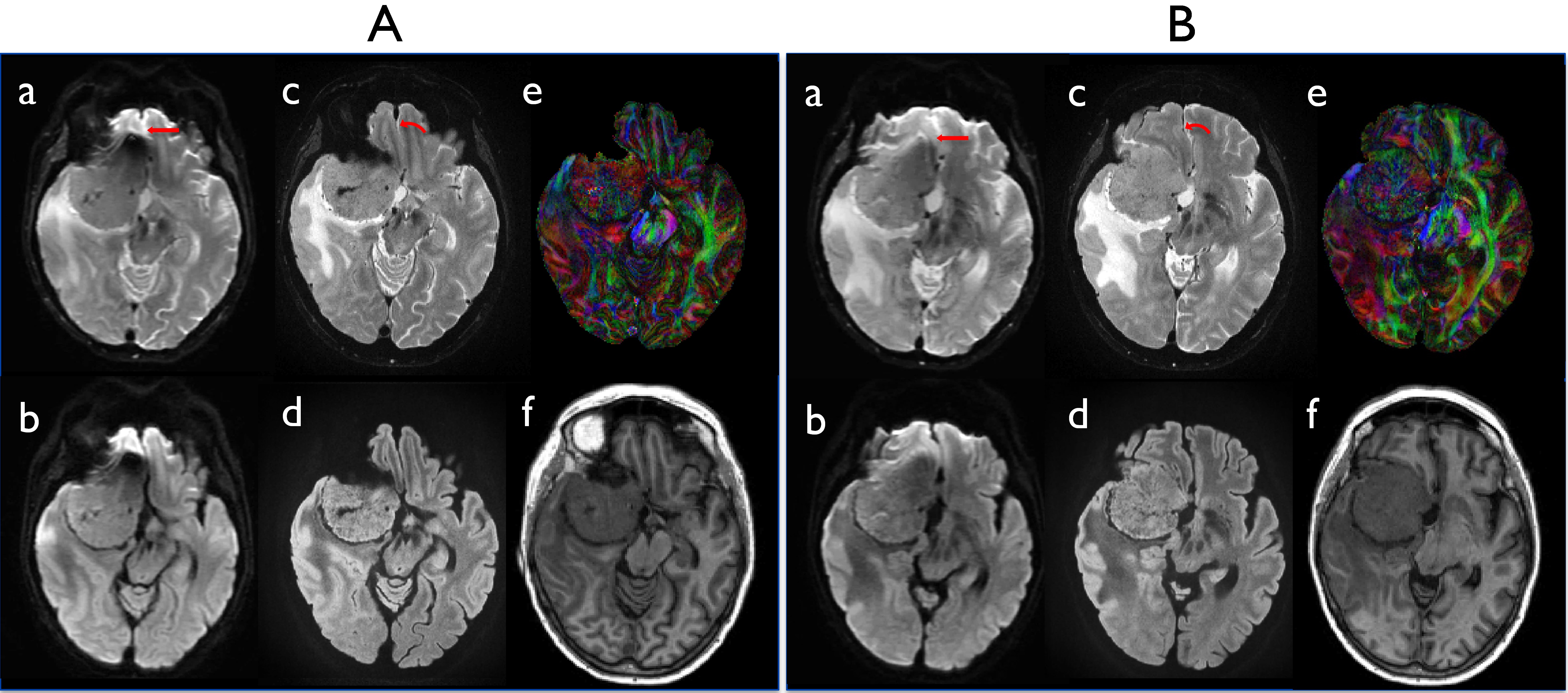
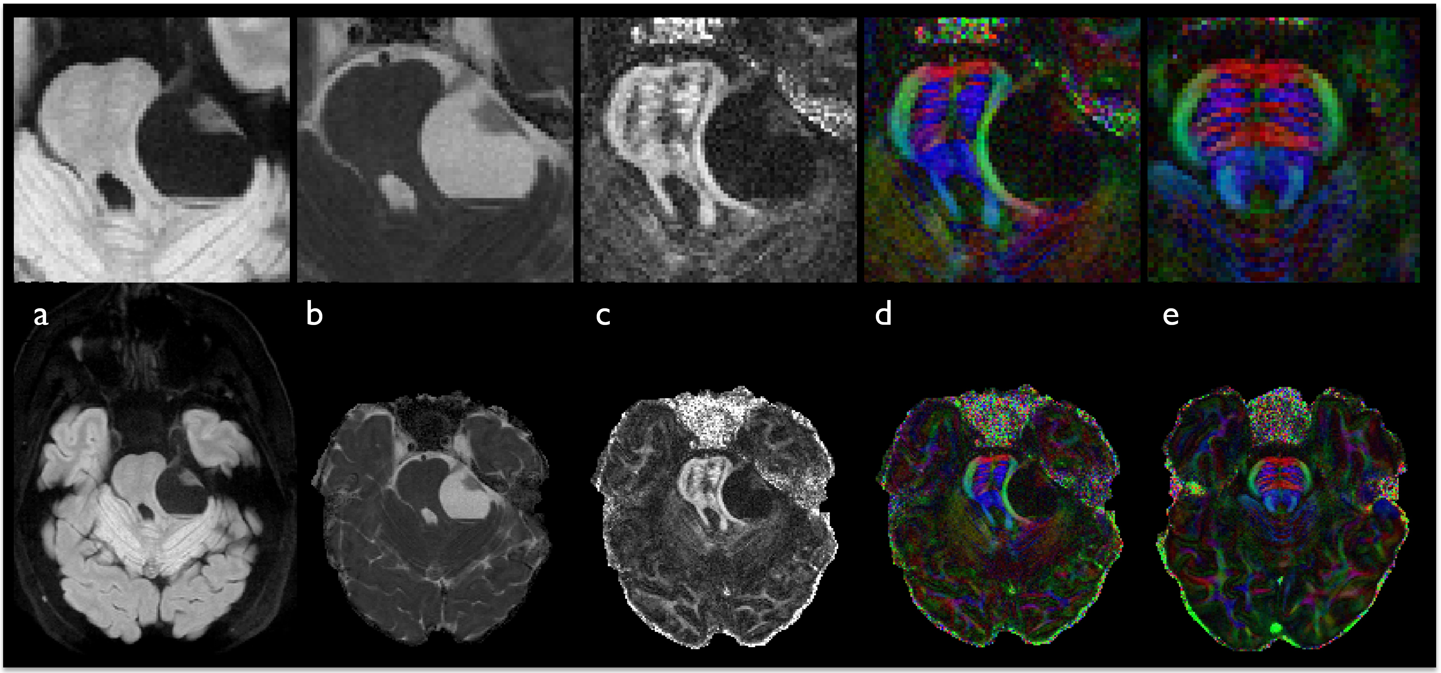
Combining a parallel imaging factor of 2 and the high gradient slew-rate available on the compact 3T resulted in a short effective echo spacing of 320 µs. However, severe distortions including image stretching and pile-up appeared in regions of high susceptibility with ssEPI-based high-resolution DWI (Fig. 1Aa-b). The spatially varying residual distortions in the areas of high susceptibility resulted in unclear boundaries between the brain tumor and the surrounding tissue. In contrast, the proposed high-resolution DIADEM provided a high degree of anatomic detail without any observable geometric distortion. This enabled a clear characterization of the brain tumor, even near high susceptibility areas adjacent to tissue-air interfaces (Figs. 1Ac-e and Bc-e). In addition, when the tumor is sufficiently large enough to cause mass effect on adjacent structures, DIADEM provides high spatial resolution diagnostic images and corresponding color-coded fractional anisotropy diffusion images in regions adjacent to the tumor (Fig. 2). Although these preliminary results on the compact 3T appear promising in the evaluation of patients with brain tumors compared to ssEPI, they are preliminary and further investigation is necessary. In order to make this approach available with a clinically feasible acquisition time on a conventional clinical scanner with standard gradients (e.g. 200 T/m/s), the combination with other approaches7,8 to further accelerate the scan time will be required.Conclusion
We have demonstrated the potential clinical use of a multi-shot method using spin-warp echo-planar encoding technique inspired by point-spread function mapping on a compact 3T scanner for diffusion imaging of brain tumors. Since this approach is not degraded by the off-resonance-induced distortions observed with ssEPI and the technique’s multi-shot nature enables high spatial resolution diffusion imaging, substantial improvements in characterizing brain tumors can be achieved in areas of high susceptibility. With the high spatial resolution capability the proposed approach can be particularly useful for evaluating brain tumors and surrounding structures occurring in regions degraded by susceptibility artifacts such as the skull base.Acknowledgements
This work was supported by NIH U01 EB024450-01.References
1. In MH, Posnansky O, Speck O. High-resolution distortion-free diffusion imaging using hybrid spin-warp and echo-planar PSF-encoding approach. Neuroimage. 2017 Mar 1;148:20-30. doi: 10.1016/j.neuroimage.2017.01.008.
2. Tan ET, Lee SK, Weavers PT, Graziani D, Piel JE, Shu Y, Huston J 3rd, Bernstein MA, Foo TK. High slew-rate head-only gradient for improving distortion in echo planar imaging: Preliminary experience. J Magn Reson Imaging. 2016 Sep;44(3):653-64. doi: 10.1002/jmri.25210.
3. Lee S-K, Mathieu J-B, Graziani D, et al. Peripheral nerve stimulation characteristics of an asymmetric head-only gradient coil compatible with a high-channel-count receiver array. Magn Reson Med 2016;76:1939–1950. doi: 10.1002/mrm.26044.
4. Weavers PT, Shu Y, Tao S, Huston J 3rd, Lee SK, Graziani D, Mathieu JB, Trzasko JD, Foo TK, Bernstein MA. Technical Note: Compact three-tesla magnetic resonance imager with high-performance gradients passes ACR image quality and acoustic noise tests. Med Phys. 2016 Mar;43(3):1259-64. doi: 10.1118/1.4941362.
5. Tao S, Weavers PT, Trzasko JD, Shu Y, Huston J 3rd, Lee SK, Frigo LM, Bernstein MA. Gradient pre-emphasis to counteract first-order concomitant fields on asymmetric MRI gradient systems. Magn Reson Med. 2017 Jun;77(6):2250-2262. doi: 10.1002/mrm.26315.
6. Weavers PT, Tao S, Trzasko JD, Frigo LM, Shu Y, Frick MA, Lee SK, Foo TKF, Bernstein MA. B0 concomitant field compensation for MRI systems employing asymmetric transverse gradient coils. Magn Reson Med. 2017 doi:10.1002/mrm.26790
7. Beuer FA, Blaimer M, Heidemann RM, Mueller MF, Griswold MA, Jakob PM. Controlled aliasing in parallel imaging results in higher acceleration (CAIPIRINHA) for multi-slice imaging. Magn Reson Med. 2005 Mar;53(3):684-91.
8. Setsompop K1, Gagoski BA, Polimeni JR, Witzel T, Wedeen VJ, Wald LL. Blipped-controlled aliasing in parallel imaging for simultaneous multislice echo planar imaging with reduced g-factor penalty. Magn Reson Med. 2012 May;67(5):1210-24. doi: 10.1002/mrm.23097.
Figures