2048
Silent Corrected Using Second Image (SCUSI) - Application of the MP2RAGE formalism to T1-weighted Zero Time Echo Imaging1GE Healthcare, Pisa, Italy, 2GE Healthcare, Munich, Germany, 3Imago7, Pisa, Italy, 4GE Healthcare, Waukesha, WI, United States
Synopsis
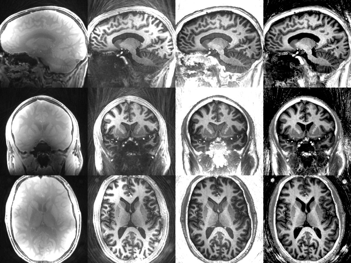
We applied the MP2RAGE formalism to a T1-weighted Zero Time Echo sequence. The complex ratio of ZTE images taken with and without inversion preparation showed a correction of the receive coil bias. Brain images of the head are presented showing improved contrast between grey and white matter.
Introduction
Providing good grey/white matter contrast in the brain using T1-weighted imaging at fields of 7T becomes more challenging due to inhomogeneity of both transmit RF (B1+) and receiver coil (B1-) fields. The application of a T1-weighted Zero Time Echo (ZTE) sequence - Silent - to brain imaging at 7T has previously been reported1 which provides acceptable SNR, strong Gray/White matter contrast, and image quality suitable for diagnostic use2, but variations in image intensity caused by B1- and B1+ are still apparent.
The MP2RAGE formalism3, in which a ratio is taken of T1-weighted MPRAGE images with and without the inversion pulse4, shows good G/W Matter contrast, and T1-weighted image uniformity which is excellent at 3T, and good at 7T. B1- inhomogeneities ("receive coil bias") are corrected, and B1+ inhomogeneities are mitigated, though both these papers show by theory and results that this correction is not perfect in inferior regions of the brain (see Marques, fig3; Van de Moortele fig 12).
We applied the MP2RAGE approach to T1-weighted ZTE. The complex ratio of images taken with and without inversion showed improved G/M contrast, and correction of B1- and B1+ inhomogeneities in the brain at 7T.
Methods
T1-weighted ZTE images were acquired on a General Electric MR 950 7T scanner (gradients=50mT/m, slew=200T/m/s; Nova Medical birdcage transmission coil, Nova Medical 32-channel receive array coil). A healthy male adult volunteer was scanned. Image parameters were similar to those previous published2 (receiver bandwidth=31.25kHz, time of inversion=600 ms, time of repetition of ZTE block=525 ms, time of echo=16 µs, flip angle (FA)=4°, spokes per segment=384, post-segment time of delay=2000 ms, cubic field of view (FOV)=192 mm; voxel size=1×1×1 mm3) but a second image was taken without the inversion pulse and with a delay time of 2600ms. RF receive and transmit parameters (transmit and receive gain, centre frequency) were held constant between the two images. Images were reconstructed in Matlab, and the simple (from the standard magnitude image reconstruction) and complex ratios (from real and imaginary components of the image reconstruction) were calculated.Results
Proton density Silent, T1-weighted Silent, and simple and complex ratio SCUSI images are displayed in fig 1. Receiver coil bias is evident in the raw proton density and T1-weighted Silent images. Visual inspection indicates an improvement in Grey/White matter contrast and image uniformity in the complex ratio images, which extends throughout the entire brain.Discussion
While the ZTE imaging module is very different, the Silent sequence in this work includes both the inversion preparation pulse and the post-acquisition delay used by MPRAGE, so to a first approximation, the relaxation equations used in MPRAGE can be applied here. This allows a good correction of coil receiver bias using the MP2RAGE approach.
In comparison with the drop-off observed in the inferior regions of the brain at 7T in the original papers attributed to failure of the inversion pulse (Van de Moortele, fig 12; Marques, fig 3), image uniformity appears to be good throughout the whole brain. This is because the Silent sequence uses an inversion pulse with a high adiabatic threshold5 which provides more consistent T1-weighting in the presence of transmit RF inhomogeneities1 found in the inferior parts of the brain.
Conclusion
The MP2RAGE approach has been applied to a ZTE sequence (Silent) to correct for receiver coil bias. The processed "SCUSI" images show improved gray/white matter contrast and image uniformity compared to the uncorrected images. These two important image quality characteristics give T1-weighted ZTE imaging increased clinical utility at 7T for diagnostic assessment and should facilitate the application of advanced image processing methods such as automated tissue segmentation.Acknowledgements
No acknowledgement found.References
1) Kelley DAC, McKinnon GC, Sacolick L et al. Optimization of a Zero Echo Time (ZTE) Sequence at 7T with Phased Array Coils. Proc ISMRM (2014) #4933
2) Costagli M, Symms MR, Angeli L et al. Assessment of Silent T1-weighted head imaging at 7 T. Eur Radiol. (2016) 26(6):1879-88
3) Marques JP, Kober T, Krueger G et al. MP2RAGE, a self bias-field corrected sequence for improved segmentation and T1-mapping at high field. Neuroimage. (2010) ;49(2):1271-81.
4) Van de Moortele P-F, Auerbach EJ, Olman C et al. T1 weighted brain images at 7 Tesla unbiased for Proton Density, T2⁎ contrast and RF coil receive B1 sensitivity with simultaneous vessel visualization. NeuroImage (2009) 46:432–446
5) Garwood M, DelaBarre L. The return of the frequency sweep: designing adiabatic pulses for contemporary NMR. J Magn Reson. (2001) 153(2):155-77.
Figures