2030
MRI Hippocampal subfield volume analysis: Comparison between Alzheimer's disease, mild cognitive impairment, and normal aging subjects in an amyloid PET project.1Department of Radiology, Faculty of Medicine Siriraj Hospital, Mahidol University, Bangkok, Thailand, 2Department of Geriatric Medicine, Faculty of Medicine Siriraj Hospital, Mahidol University, Bangkok, Thailand
Synopsis
Hippocampal atrophy evidenced by MRI is one of the most validated biomarkers of Alzheimer’s disease (AD). The previous neuropathological data showed a differential vulnerability of hippocampal subfields to AD processes. This study aims to use an automated analysis technique for subfield hippocampal volume measurement in order to differentiate early detection of AD. We demonstrated high diagnostic efficacy of using hippocampal subfield analysis for discriminating AD subjects from heathy control (HC) or mild cognitive impairment (MCI) than whole hippocampal volume and feasibility for discriminating MCI to HC as compared with amyloid PET result.
Introduction:
Hippocampal atrophy evidenced by MRI is one of the most validated biomarkers of AD. Based on neuropathological data showing a differential vulnerability of hippocampal subfields to AD processes, hippocampal subfield analysis could improve the diagnostic accuracy of AD or MCI 1 . In this study, we aimed to demonstrate the difference of hippocampal subfield volumes by using automated subfield volumetric analysis for discriminating between AD, MCI and HC.Material and methods:
A total 53 age-matched subjects included 15 AD patients (mean 72.3±6.9 years), 9 MCI patients (mean 68.4±3.9 years) and 29 HC subjects (mean 69.5±5.1 years) were included in this study with analysis of neurological and neuropsychiatric test and underwent MRI (3-Tesla) with high-resolution 3D-T1W covering the whole brain(FOV 230 × 230 × 172 mm3, matrix size 352x352, voxel size 0.72 × 0.72 × 0.65 mm3, TE /TR 4.8/9.8 ms; FA 8°, scan time 6 min). Quantitative volumetric analysis of hippocampal subfields was performed by using Freesurfer (v. 6.0) 2 for comparing AD, MCI and HC subjects.Results:
There was a widespread pattern of subfield atrophy. Right subiculum, right presubiculum, bilateral molecular layer, and bilateral fimbria volumes were higher diagnostic efficacy than whole hippocampal volume for discriminating AD subjects from HC or MCI subjects. Only left fimbria volume was significantly reduced in MCI subjects compared to HC (normalized volume 47.7, sensitivity = 66.7%, specificity = 89.7%, AUC 0.709). The accuracy of left fimbria as the predictor of MCI converter was 75% when compared with amyloid PET result.Conclusion:
The structural brain imaging using quantitative volumetric analysis of hippocampal subfield could be used for differentiating AD, MCI and HC. These findings may be helpful for early detection and follow up of AD and MCI patients in the clinical setting.Acknowledgements
The authors thank Mrs. Angkana Jongsawaddipatana and all staff members of the primary care unit of Siriraj Hospital for their contribution to data collection.References
1. De Flores R, La Joie R, Chételat G. Structural imaging of hippocampal subfields in healthy aging and Alzheimer’s disease. Neuroscience. 2015;309:29–50.
2. Iglesias JE, Augustinack JC, Nguyen K, Player CM, Player A, Wright M, Roy N, Frosch MP, McKee AC, Wald LL, Fischl B, Van Leemput K, Alzheimer's Disease Neuroimaging Initiative (2015) A computational atlas of the hippocampal formation using ex vivo, ultra-high resolution MRI: application to adaptive segmentation of in vivo MRI. Neuroimage 115:117–137
Figures
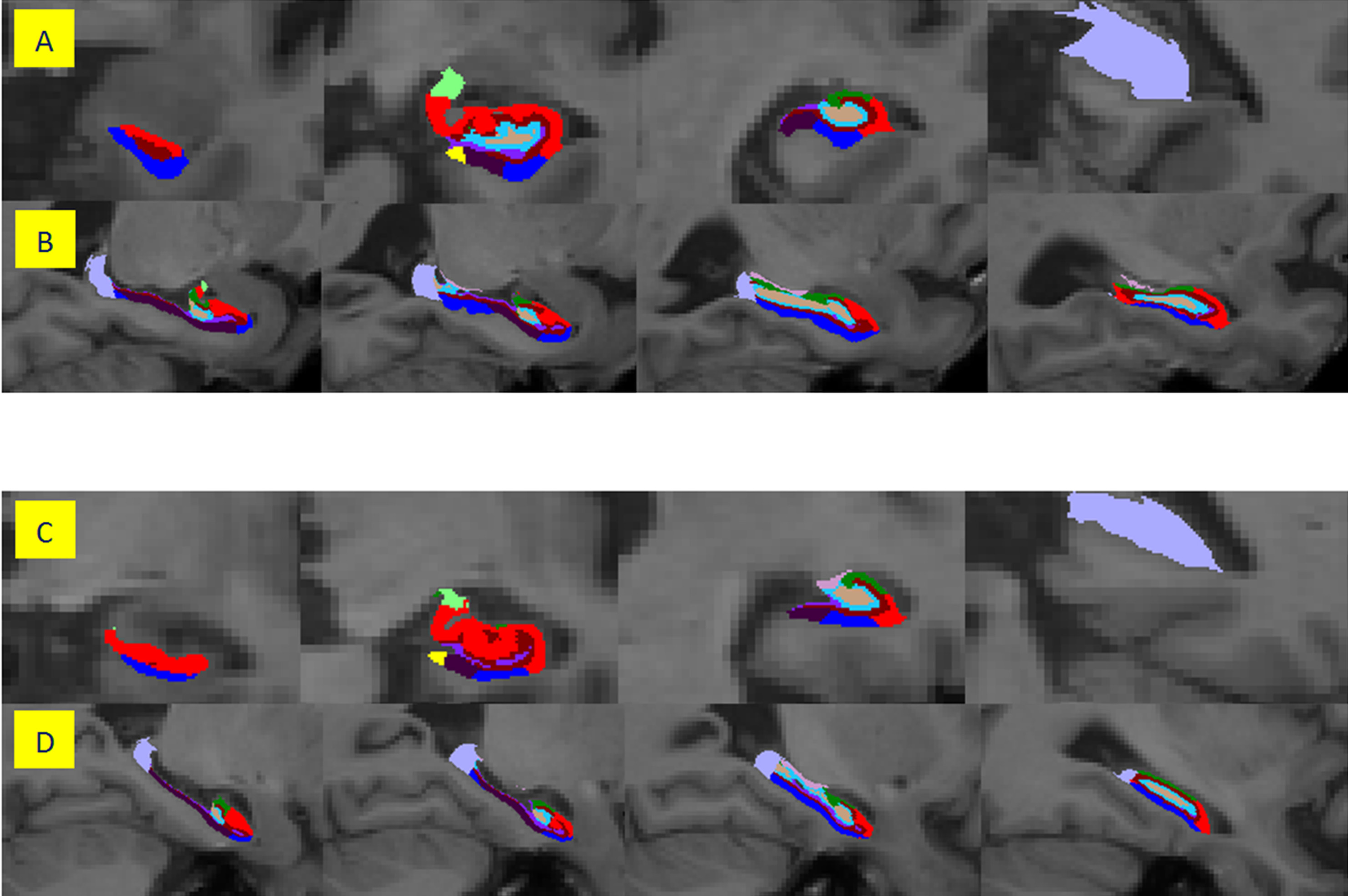
Figure 1: A Coronal slices from anterior (left) to posterior (right) and B sagittal slices from medial (left) to lateral (right) of a 65-year-old man, healthy control. C Coronal slices from anterior (left) to posterior (right) and D sagittal slices from medial (left) to lateral (right) of a 71-year-old female diagnosed Alzheimer’s disease