2026
Changes in Functional and Structural Brain Connectome Along the Alzheimer’s Disease ContinuumFederica Agosta1, Silvia Basaia1, Elisa Canu1, Francesca Imperiale1, Giuseppe Magnani2, Monica Falautano2, Giancarlo Comi2, Andrea Falini3, and Massimo Filippi1,2
1Neuroimaging Research Unit, INSPE, Division of Neuroscience, San Raffaele Scientific Institute, Vita-Salute San Raffaele University, Milan, Italy, 2Department of Neurology, San Raffaele Scientific Institute, Vita-Salute San Raffaele University, Milan, Italy, 3Department of Neuroradiology, San Raffaele Scientific Institute, Vita-Salute San Raffaele University, Milan, Italy
Synopsis
We investigated structural and functional brain network architecture in patients with Alzheimer’s disease (AD) and mild cognitive impairment (MCI); and assessed the relationship between healthy brain network functional connectivity and the topography of brain atrophy in patients along the AD continuum. Structural connectivity alterations distinguished MCI who converted to AD from those who did not. Brain regions most strongly connected with the disease-epicenter (left hippocampus) in the healthy functional connectome were also the most atrophic in both AD and converters MCI. Graph theoretical analysis provides insight on how neurodegeneration propagates across the human brain in the early phase of AD.
Introduction
Abnormal propagation of amyloid β and tau in Alzheimer’s disease (AD) has been suggested to be mediated by the extremely complex, yet highly structured topology of the underlying connectional architecture. The study of the effects of the disease on the brain networks along the AD continuum is critical in conceptualizing and predicting this networked disease evolution in vivo. In this study, we investigated structural and functional brain network architecture in patients with AD and amnestic mild cognitive impairment (aMCI), stratified in converters (c-aMCI) and non-converters (nc-aMCI) to AD; and assessed the relationship between healthy brain network functional connectivity and the topography of brain atrophy in patients along the AD continuum.Methods
Ninety-four AD patients, 47 aMCI patients (25 c-aMCI within 36 months) and 53 age- and sex-matched healthy controls were studied. Participants underwent 3D T1-weighted MRI, diffusion tensor MRI and resting state functional MRI. Graph analysis and connectomics assessed global and local, structural and functional topological network properties and regional connectivity. Healthy topological features of brain regions were assessed based on their connectivity with the point of maximal atrophy (epicenter) in AD and aMCI patients.Results
Brain network graph analysis properties were severely altered in AD patients. Structural brain network was already altered in c-aMCI patients relative to healthy controls in particular in the temporal and parietal brain regions, while functional connectivity did not change. Structural connectivity alterations distinguished c-aMCI from nc-aMCI cases. In both AD and c-aMCI, the point of maximal atrophy was located in left hippocampus (epicenter). The brain regions with the strongest functional connectivity strength and the shortest topological distance with the epicenter in healthy subjects included the right posterior cingulate cortex and hippocampus, left orbitofrontal gyrus and bilateral temporal pole, parahippocampi, amygdala and precuneus. In AD and c-aMCI, regional atrophy correlated negatively with the regional topological distance with the epicenter in controls (Figure). Furthermore, a positive correlation was also found between regional atrophy in patients and the functional connectivity strength with the epicenter in controls (Figure).Discussion
Progressive degeneration in the AD continuum is associated with an early breakdown of anatomical brain connections and follows the strongest connections with the disease-epicenter. These findings support the hypothesis that the topography of brain connectional architecture can modulate the spread of AD through the brain.Conclusions
Characterizing the brain as a network using diffusion tensor and resting state functional MRI and graph theoretical analysis provides insight on how neurodegeneration propagates across the human brain in the early phase of AD.Acknowledgements
The study was supported by the Italian Ministry of Health (grant number GR-2010-2303035) and Alzheimer’s Drug Discovery Foundation (grant number 20131211).References
No reference found.Figures
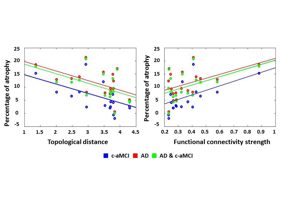
Figure: Scatterplots of the correlations between regions of interest (ROI) percentage of atrophy in AD and c-aMCI patients and
ROI topological distance from and functional connectivity strength with the
epicenter (hippocampus) in independent healthy controls. ROIs are the 20%
highest scoring ROIs according to the sum of their rank position in functional
connectivity strength (highest) with and topological distance (lowest) from the
disease-epicenter in independent healthy controls. Abbreviations: AD= Alzheimer’s Disease; aMCI= amnestic mild
cognitive impairment (c= converters).