2023
Correlation analysis between the gray matter volumes obtained with two different imaging sequences and the cognitive decline in Apolipoprotein E ε4 carrier subjects1Radiology, Kyung Hee Univ. Hospital at Gangdong, Seoul, Republic of Korea, 2Neurology, Kyung Hee Univ. Hospital at Gangdong, Seoul, Republic of Korea
Synopsis
To evaluate the association between GMV loss and cognitive decline in the APOE e4 carriers and to investigate alterations of GMV, MPRAGE and DIR images were acquired from 72 subjects (51 noncarriers, 21 carriers). Voxel- and ROI-based analyses were performed to evaluate the association between GMV loss and the MMSE score and to do the group differences of GMV for each sequence. GMV of carriers was positively correlated with the MMSE score for both sequences. DIR can be effective for identifying GMV loss in the carriers and may be useful to evaluate GMV changes in the early stage of dementia.
Background
The ε4 allele of the apolipoprotein E (APOE) gene is the major genetic risk factor for Alzheimer’s disease. Structural MRI, especially 3D T1W image is used as a gold standard to evaluate brain atrophy in AD by using voxel-based morphometry (VBM). However, another recent study of ours suggests the DIR images as the new imaging biomarker to evaluate GMV loss in AD patients are more sensitively (1). Previous studies showed GMV loss on T1WI in the carriers compared with the non-carriers (2, 3). However, there is no study to evaluate the association between GMV changes and cognitive impairment in APOE ε4 carriers. The purposes of this study are to evaluate the association between GMV loss and cognitive decline in the carriers using both DIR and T1W images and to investigate alterations of GMV between 2 groups using both sequences.Materials and Methods
Seventy-two subjects (51 noncarriers: 66.0 ± 8.7 years, 21 carriers:70.6 ± 5.8 years) were prospectively enrolled. The cognitive function was assessed by Korean version of the Mini-Mental State Examination (K-MMSE). The K-MMSE scores were 24.9 ± 5.6 for noncarriers and 19.9 ± 6.6 for carriers. All MR images were acquired with using a 3 tesla MR system. We performed the T1W MPRAGE image with an isotropic voxel size of 1 mm3. In a DIR sequence, we adjusted the inversion times to suppress CSF (TI1 of 2930 ms) and white matter (TI2 of 350 ms) signals. The detailed information for the DIR sequence was published previously (Jahng GH. et al. 2016). SPM8 software was used to perform brain tissue segmentation, creating template using DARTEL, normalization and smoothing. To evaluate the association between GMV loss and the K-MMSE score for each sequence, voxel-based multiple regression analyses with regress-out of age and gender were performed. Furthermore, to compare GMVs between carriers and non-carriers for each sequence, voxel-based two sample t-test was used. P-value of 0.0001 without multiple comparisons with cluster size of 20 was used. Finally, we obtained the region-of-interests (ROIs) based on the results of the VBM analysis. We also conducted ROI-based correlation analysis, to evaluate the association between GMV loss and the K-MMSE score for each sequence. Furthermore, to compare GMVs between carriers and non-carriers for each sequence, ROI-based two sample t-test was used. P-value of 0.05 was used for the ROI analysis.Results
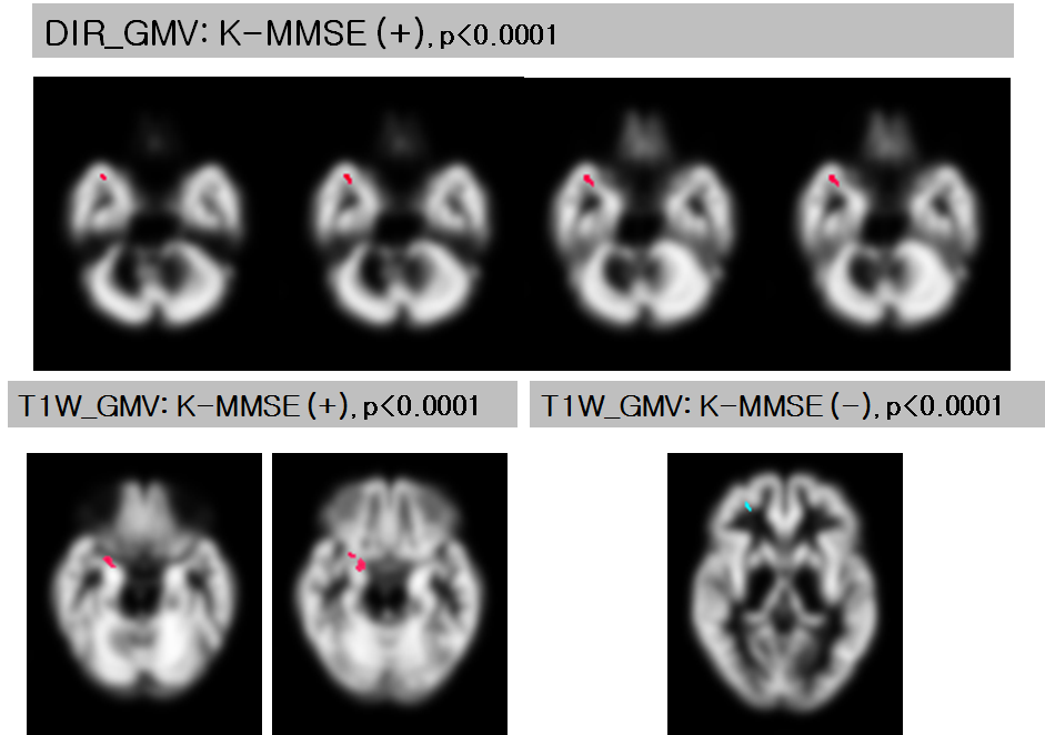
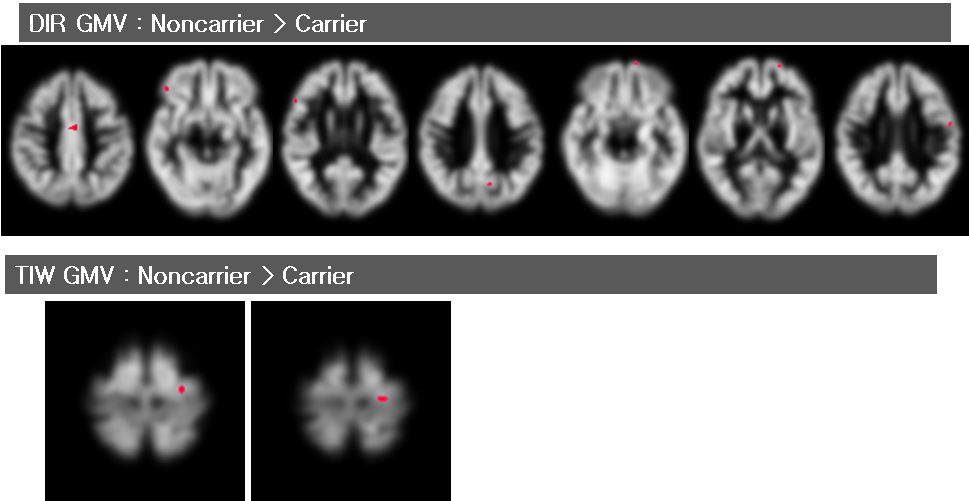
In the voxel-based multiple regression analysis, GMV on DIR images in the carrier group was positively correlated with the K-MMSE score within a region of right superior temporal gyrus. On T1WI, the GMV of carriers was positively correlated with the K-MMSE score within the right frontal gyrus and also, a negative correlation was observed within the right medial frontal gyrus. In the ROI-based analysis, only on DIR, the GMV was positively correlated with the K-MMSE score within the right middle frontal gyrus in the non-carriers. In the carrier group, the ROI-based correlation analysis showed that the GMV was positively correlated with the K-MMSE score within the right inferior frontal gyrus on both T1W and DIR images. There is a significantly negative correlation between GMV and MMSE score on TIWI, which is an unusual result. In voxel-based comparison of the GMV, the carrier group had a smaller GMV on DIR. On T1W, the GMV in carriers was decreased compared with noncarrier. On the ROI-based analysis, GMV on T1W was significantly different between the two groups at the left precentral and postcentral gyri, but not on DIR. In contrast, GMV on DIR was significantly different between the two groups at the left cingulate gyrus, and the right middle frontal gyrus, but not on T1W. For left amygdala, only the DIR showed a significant GMV loss.Discussion
GMV with T1W has the inconsistent result. DIR is sensitive to measure the association between GMV and the cognitive function in the non-carriers as well as in carriers. Both DIR and T1W showed higher GMV in the non-carrier than that of the carrier. T1W and DIR sequences revealed different regions with significant GMV loss. However, there were more regions with significant GMV loss on DIR, and especially in the left amygdala, the carriers showed a significant GMV loss.Conclusion
DIR is sensitive to evaluate the association between GMV and the cognitive function in the non-carriers as well as in the carriers. Only on DIR sequence, GMV loss was significant at the amygdala in the APOE ε4 carriers. DIR can be effective for identifying GMV loss in the carriers and may be useful to evaluate GMV changes in the early stage of dementia.Acknowledgements
The research was supported by the Basic Science Research Program through the National Research Foundation of Korea (NRF) grant funded by the Korea government (MSIP) (2014R1A2A2A01002728) and by the Convergence of Conventional Medicine and Traditional Korean Medicine R&D program funded by the Ministry of Health & Welfare through the Korea Health Industry Development Institute (KHIDI) (HI16C2352).References
1. Jahng GH, et al. Double inversion recovery imaging improves the evaluation of gray matter volume losses in patients with Alzheimer's disease and mild cognitive impairment. Brain Imaging Behav. 2016;10(4):1015-1028.
2. Filippini N, et al. Anatomically-distinct genetic associations of APOE epsilon4 allele load with regional cortical atrophy in Alzheimer's disease. Neuroimage. 2009; 1;44(3):724-8.
3. Liu Y, et al. Effect of APOE ε4 allele on cortical thicknesses and volumes: the AddNeuroMed study. J Alzheimers Dis. 2010;21(3):947-66.
Figures