2021
Increased Mode of Anisotropy in crossing-fibre areas predicts conversion from Mild Cognitive Impairment (MCI) to Alzheimer's disease (AD)1Clinical Imaging Sciences Centre, Brighton and Sussex Medical School, Brighton, United Kingdom, 2School of Life Sciences, University of Sussex, Falmer, United Kingdom, 3Neuroimaging Laboratory, Santa Lucia Foundation IRCCS, Rome, Italy
Synopsis
Diffusion MRI was used to examine whether any change in the white matter tracts of patients with mild cognitive impairment (MCI) can predict conversion to Alzheimer’s disease (AD) in a longitudinal study. Our data show increases in mode of anisotropy (MO) in a region of crossing fibres in the centrum semiovale for MCI patients who later converted to AD.
Background
Mild cognitive impairment (MCI) is associated with an increased risk for developing dementia in a short time period1. Nevertheless, MCI is a heterogeneous condition, and valid biomarkers able to predict conversion are desperately needed. MRI is sensitive to different aspects of tissue modifications across Alzheimer’s disease (AD) in both white and grey matter2. A previous study extensively compared indices derived from diffusion tensor image (DTI) across AD, MCI and age-matched controls3. While significant changes in all indices were found in AD compared to the other groups, the only parameter that was significantly different between MCI and healthy controls was the mode of anisotropy (MO), which quantifies the shape of the diffusion tensor. The aim of this longitudinal study is to assess whether any index derived from DTI is able to predict conversion from MCI to AD.Methods
Data from 54 MCI patients (M/F=29/25, mean age = 69.8 years (SD 7.77), range: 47-82 years) were acquired at 3T. The protocol included a T1-weighted volume, DTI and resting state functional MRI. Patients were reassessed after a period of 24 months (± 6 months), and AD conversion recorded. Patients were thus classified as MCI converters and non-converters.
All diffusion-weighted images were corrected for involuntary motion and eddy current distortions using affine registration and the FMRIB's Linear Registration Tool (FLIRT). The images were skull-stripped using FMRIB's Brain Extraction Tool (BET), and FMRIB’s dtifit was used to fit tensor indices: Fractional Anisotropy (FA), Axial Diffusivity, Radial Diffusivity, Mean Diffusivity and Mode of Anisotropy (MO). The FA data were then warped into MNI space using the Advanced Normalization Tools (ANTs) 2.1.0., and the resultant warpings were applied to the remaining tensor indices.
Voxel-wise analysis was performed using Tract-Based Spatial Statistics (TBSS). Permutation inference4 was performed on the skeletonised images with randomise, available with FSL, using the Threshold-Free Cluster Enhancement (TFCE) method. Results were accepted as significant for p < 0.05 after correction for multiple comparisons.
Results
At follow-up, 12 patients (M/F=4/8, mean age = 73.1 years (SD 5.60), range: 63-82 years) had converted to AD, while 42 patients (M/F=25/17, mean age = 68.8 years (SD 8.10), range: 47-81 years) remained as MCI.
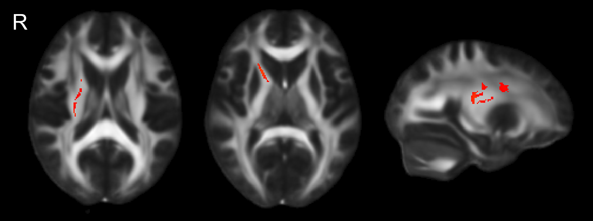
Increased MO was found in a region of crossing fibres in the right centrum semiovale and internal capsule (Fig 1). No other tensor indices showed significant differences between MCI converters and non-converters.
Discussion
Our data confirm that MO is extremely sensitive to early changes occurring in the white matter of individuals with MCI. Remarkably, changes to this parameter were able to differentiate between patients who converted to AD within 2 years and those who did not. The location of the MO increase, within a region of the corona radiata where the superior longitudinal fasciculus crosses the anterior thalamic radiation and the corticospinal tract, matches the findings of Douaud et al3. Above and beyond their findings of increased MO in MCI compared to healthy controls, we show here that increased MO can differentiate between MCI converters and non-converters at baseline, when these individuals are indistinguishable from a clinical and neuropsychological perspective. Following the interpretation given in 3, which was based on further tractographic analysis, we speculate that selective degeneration of the superior longitudinal fasciculus with a relative preservation of the corticospinal tract could cause the observed MO changes. We are now in the process of correlating these findings with changes in neuropsychological scores, and to establish the value of this biomarker at single-subject level.Acknowledgements
MC Gabel is funded by the Motor Neurone Disease Association.References
- Manly JJ, Tang MX, Schupf N, et al. Frequency and Course of Mild Cognitive Impairment in a Multiethnic Community. Ann Neurol, 2008, 63(4): 494-506
- Jack CR Jr, Knopman DS, Jaqust WJ, et al. Tracking pathophysiological processes in Alzheimer's disease: an updated hypothetical model of dynamic biomarkers. Lancet Neurol. 2013, 12(2):207-16
- Douaud G, Jbabdi S, Behrens TE, et al. DTI measures in crossing-fibre areas: Increased diffusion anisotropy reveals early white matter alteration in MCI and mild Alzheimer's disease. NeuroImage. 2011, 55:880-890
- Winkler AM, Ridgway GR, Webster MA, et al., Permutation inference for the general linear model, NeuroImage, 2014, 92:381-397
Figures