2018
Follow up research of hippocampal subfield in patients with mild Alzheimer's diseaseYing Liu1 and Lizhi Xie2
1Radiology Department, Peking University Third Hospital, Beijing, China, 2GE Healthcare, China, Beijing, China
Synopsis
The aim of this study is to evaluate the atrophy pattern of hippocampal subfield and follow up the changes of hippocampal subfield by using automatic segmentation tool in patients with mild AD. The results indicate that volumes of hippocampal subfield decrease in patients with mild AD, and the declination are positive correlated with clinical scores. We conclude that substructures of hippocampal might serve as a good index to characterize subtle changes in AD patients.
Purpose
Alzheimer’s disease (AD) is characterized by the progression of cognitive and behavioral changes. Using magnetic resonance imaging (MRI), brain atrophy has been detected in certain cerebral areas, especially in hippocampus [1], in AD patients. Some studies focused on hippocampal subfield [2-3]. The aim of this study is to evaluate the atrophy pattern of hippocampal subfield and follow up the changes of hippocampal subfield by using automatic segmentation tool in patients with mild AD.Material and Methods
Eleven patients with AD (4 females, 64-86 years, mean age = 75.50±7.04 years) were recruited in this study. For comparison, ten healthy controls (5 females, age range 60-77 years, mean age 69.50±6.91 years) were also included. MMSE scores and CASI scores were measured in all subjects. MRI examinations were performed using a 3.0T scanner. The acquisition parameters were as follows: for T1-weighted imaging, TR = 2530ms, TE = 3.44ms, flip angle = 7°, slice thickness = 1 mm, voxel size = 1.0×1.0×1.0 mm3, scan time = 6 min 3 sec; for coronal T2-weigthed imaging, TR = 4140ms, TE = 97ms, slice thickness = 2 mm, voxel size = 0.6×0.5×2.0 mm3, scan time = 5 min 20 sec. Automatic Segmentation of Hippocampal Subfields software (ASHS, http://www.nitrc.org/projects/ashs/) was used to analyze the acquired anatomical images. The differences of the two groups were assessed by performing two-sample t test. Correlations between the scores of MMSE and volumes of hippocampal subfield were explored using Pearson’s correlation analysis. Correlations between the scores of CASI and volumes of hippocampal subfield were explored using Pearson’s correlation analysis. Seven of the eleven AD patients were scanned twice. The average scanning interval was 14.40±2.95 months.Results
1. In patients with mild AD, mean MMSE scores were (23.40±1.78), mean CASI scores were (82.70±5.32), mean years of education were (13.70±4.67). In healthy controls, mean MMSE scores were (29.38±0.52), mean CASI scores were (97.63±1.60), mean years of education were (15.38±1.41). MMSE scores and CASI scores were significantly different between the patient and control groups (p=0.000), especially in certain subitems of CASI scores, like short-term memory (p=0.000), focus (p=0.016), direction (p=0.003), and thinking (p=0.025). 2. Compared to the healthy controls, we found significant decrease in volumes of hippocampal subfields in patients with mild AD in bilateral Cornu Ammomis 1 (CA1) (L: p=0.025, R: p=0.001), bilateral subculum (SUB) (L: p=0.005, R :p<0.001), bilateral dentate gyrus (DG) (L: p=0.048, R: p=0.004), bilateral perirhinal cortex (L: p=0.022, R: p=0.001), right CA2 (p=0.011), and right entorhinal cortex (ERC) (p=0.005). There was no significant difference in intracranial volume (ICV) (p>0.05) (Figure1). 3. There were positive correlations between volumes of right CA1, right SUB, right ERC, right perirhinal cortex and MMSE scores. There were positive correlations between volumes of left SUB, right CA1, right DG, right SUB, right perirhinal cortex and CASI scores (Table1). One of the subitems of CASI scores, long-term memory, was also found positive correlated with certain areas, especially with left DG. In addition, short-term memory scores were related with bilateral CA1, right CA2, right DG, right SUB and right perirhinal cortex; direction function were related with bilateral CA1 and bilateral SUB; and thinking were related with bilateral CA1, bilateral SUB and bilateral perirhinal cortex (Table2). 4. Compared to the volumes of hippocampal subfields from the first MRI sessions, we found significant decreasing volumes of the second sessions in the following regions: ICV (p=0.039), bilateral DG (L: p=0.011, R: p=0.043) and left SUB (p=0.034).Conclusion
Volumes of hippocampal subfield decreased in patients with mild AD and their declinations were positive correlated with clinical scores. MMSE scores and CASI scores were related to the decreases of hippocampal subfield, especially subitems of CASI scores. The short-term memory had correlations with more hippocampal subfields, which had been proved to be related to hippocampus functions. Volumes of hippocampal subfield decreased in AD patients as the disease progressed. As a result, we think substructures of hippocampal might serve as a good index to characterize subtle changes in AD patients.Acknowledgements
No acknowledgement found.References
[1] Jack CR Jr, Shiung MM, Gunter JL, et al. Comparison of different MRI brain atrophy rate measures with clinical disease progression in AD. Neurology 2004; 62(4):591-600. [2] Yushikevich PA, Wang H, Pluta J, et al.Nearly automatic segmentation of hippocampal subfields in in vivo focal T2-weighted MRI. Neuroimaage 2010;53(4):1208-1224. [3] Pluta J, Yushkevich P, Das S, et al. In vivo Analysis of Hippocampal Subfield Atrophy in Mild Cognitive Impairment via Semi-Automatic Segmentation of T2-Weighted MRI. J Alzheimers Dis 2012; 31(1):85-99.Figures
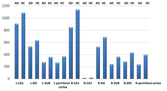
Figure.1 Significant decreased volumes of
hippocampal subfields in patients with mild AD were found in bilateral
CA1,SUB,DG,perirhinal cortex, right CA2 and right entorhinal cortex.
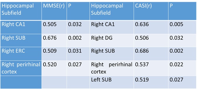
Table.1 There were positive correlations between volumes of hippocampal
subfields and MMSE scores and CASI scores.
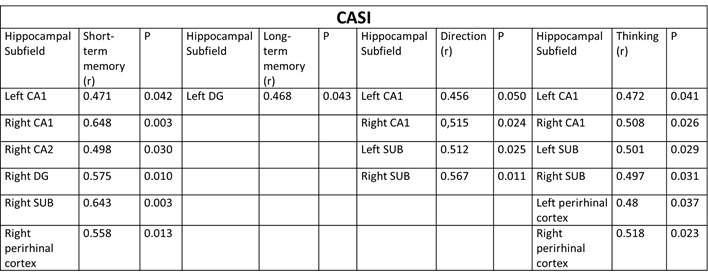
Table.2 Subitems of CASI scores had positive correlation with certain areas.
Bilateral CA1, right CA2, DG, SUB and perirhinal cortex were related with
short-term memory. Left DG was related with long-term memory. Bilateral CA1 and
SUB were related with direction function. Bilateral CA1, SUB and perirhinal
cortex were related with thinking.