1977
Comparison of Two Methods for the Measurement of T1 Hyperintensity in Multiple Sclerosis Patients with Repeated Exposure to Gadolinium-Based Contrast Agents1MS/MRI Research Group, Division of Neurology, University of British Columbia, Vancouver, BC, Canada, 2Dept of Radiology, University of British Columbia, Vancouver, BC, Canada
Synopsis
Exposure to gadolinium-based contrast agents is associated with long-term increase in T1 signal intensity in deep grey brain structures, but the measurement methodologies have not been well investigated. We propose marking regions of interest (ROIs) on registered serial T2w images, and compared two methods for measuring the signal changes in the corresponding T1w images: 1) Align the T1w to the T2w images (T2-space), and 2) Map the ROIs marked on the T2w images to the T1w images (T1-space). Applying these methods to frequent and infrequent scanning cohorts, we found signal increase to be associated with GBCA exposure, and T1-space is more sensitive.
Introduction
Gadolinium-based contrast agents (GBCA) have been widely used for magnetic resonance imaging (MRI)1,4. However, recent studies have observed an increase in T1 signal intensity in deep grey brain structures of patients receiving GBCA related to gadolinium deposition1-4. To test these observations, precise and sensitive methods for measuring signal intensity changes are needed. In most previous studies1-3, mean signal intensities were measured directly from brain regions of interest (ROIs) identified on unregistered T1-weighted (T1w) images. In contrast, because many of the deep grey brain structures are better visualized on T2-weighted (T2w) images, we propose employing groupwise registration of the T2w images to bring all time points to a common space for analysis. Accordingly, our study aims to compare two different mapping methods in their ability to detect, over time, signal intensity changes from repeated GBCA exposure.Methods
MRIs from a cohort of 200 patients, enrolled in a 2-year placebo-controlled (negative) trial assessing the efficacy of MBP82985, were acquired with a standardized MRI protocol from 47 sites using 1.0 to 3.0T scanners. 80 patients received 10 GBCA injections between baseline and year 2 (frequent cohort). The remaining 120 patients received 3 GBCA injections over the same period (infrequent cohort).
To facilitate the identification of the deep brain grey matter ROIs, groupwise registration was performed on T2w images for all patients across 3 timepoints (baseline, year 1, year 2). The ROIs were placed at the centre of the following structures: dentate (DN), globus pallidus (GP), caudate (CD), thalamus (TH), pons, and white matter (WM).
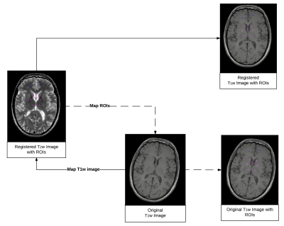
Two methods were compared for obtaining the mean signal intensity of each ROI. The first mapped the unenhanced T1w images onto the groupwise-registered T2w images. The second mapped the ROIs onto the non-registered, original unenhanced T1w images. We refer to the first and second methods as “T2-space” and “T1-space” respectively (Figure 1). Subsequently, the signal intensity of each ROI on the T1w images for both methods was measured1-3. Normalization of the signal intensity was then performed by using the pons and WM as reference standards1 (i.e. DN/pons, GP/WM, CD/WM and TH/WM), because the pons and WM have the least gadolinium accumulation, consistent with methods used in previous literature.
Statistical analyses were performed on the measurements at baseline and year 2 to determine (i) if there was an increase in T1w signal intensity over time using paired Student’s t-test, (ii) if there were differences between the frequent and infrequent cohorts using Wilcoxon rank-sum test, and (iii) the agreement between the 2 methods using Spearman’s rank correlation.
Results
For the frequent cohort (Table 1), all ROIs measured in T1-space showed increased signal intensity from baseline to year 2 (p < 0.05), while only DN and GP measured in T2-space showed a significant increase. For the infrequent cohort, only DN displayed an increase in signal intensity for both T1-space and T2-space from baseline to year 2. Comparing the frequent and infrequent cohorts in their net intensity changes from baseline to year 2, T1-space revealed a difference between the 2 cohorts in 3 of the 4 structures (GP, CD, and TH), while T2-space showed a difference in only 1 structure (GP) (Table 2). Lastly, the Spearman’s correlation for normalized ROI intensity between T1-space and T2-space in the frequent cohort was 0.90 and in the infrequent cohort was 0.82.Discussion
Our results showed an increase in T1 signal intensity in all brain structures associated with GBCA exposure1-4. DN had the largest increase over two years for both the frequent and infrequent cohorts and therefore may be the most sensitive structure for GBCA accumulation. CD, GP and TH only showed a significant increase in the frequent cohort confirming the dose-relationship for gadolinium deposition. T1-space analysis appeared to be more sensitive for detecting changes in T1 signal intensity compared to registration to T2-space. We hypothesize that registering the T1w images to T2-space blurred the intensities sufficiently to reduce sensitivity for detecting GBCA-related signal increases. Conversely, mapping the ROIs onto the original T1w images avoided altering the T1w intensities, while taking advantage of the registered T2-space for more consistent ROI identification.Conclusion
Increase in T1 signal intensity in deep grey matter from gadolinium deposition is associated the amount of GBCA exposure. T1-space method has higher sensitivity for detecting these increases.Acknowledgements
We gratefully acknowledge support from Mr Jefferson J. Mooney, A&W, the UBC MS/NMO Program, the Natural Sciences and Engineering Research Council of Canada, the MS Society of Canada, and the Maureen and Milan Ilich Foundation.References
1. Kanda T, Ishii K, Kawaguchi H, Kitajima K, Takenaka D. High Signal Intensity in the Dentate Nucleus and Globus Pallidus on Unenhanced T1-weighted MR Images: Relationship with Increasing Cumulative Dose of a Gadolinium-based Contrast Material. Radiology. 2014;270(3):834-841.
2. McDonald RJ, McDonald JS, Kallmes DF, et al. Intracranial Gadolinium Deposition after Contrast-enhanced MR Imaging. Radiology. 2015;275(3):772-782.
3. Murata N, Gonzalez-Cuyar LF, Murata K, et al. Macrocyclic and Other Non–Group 1 Gadolinium Contrast Agents Deposit Low Levels of Gadolinium in Brain and Bone Tissue. Invest Radiol. 2016;51(7):447-453.
4. Ramalho J, Semelka RC, Ramalho M, Nunes RH, AlObaidy M, Castillo M. Gadolinium-based contrast agent accumulation and toxicity: An update. Am J Neuroradiol. 2016;37(7):1192-1198.
5. Freedman MS, Bar-Or A, Oger J, et al. A phase III study evaluating the efficacy and safety of MBP8298 in secondary progressive MS. Neurology. 2011;77(16):1551-1560.
Figures


