1971
Regional cerebral blood flow alterations in patients with comitant exotropia: a pilot 3D-pCASL MRI studyZhi Wen1, Xuefang Lu1, Xin Huang2, Yang Fan3, Yunfei Zha1, and Baojun Xie1
1Dept. Radiology, Renmin Hospital of Wuhan University, Wuhan, China, 2Dept. Ophthalmology, Renmin Hospital of Wuhan University, Wuhan, China, 3GE Healthcare, Beijing, China
Synopsis
Strabismus is a common eye disease characterized by abnormal eye position and ocular motor disorder. In this study, we compared the cerebral blood flow (CBF) in patients with comitant extropia (CE) relative to healthy controls using 3D-pCASL MRI. We found that CE patients had significantly increased CBF in the right parahippocampal region, bilateral medial FG/ACC, bilateral IFG, left SFG, bilateral MCC, right MFG (BA8), and right paracentral lobule. This study demonstrates the hypothesis that CE involves the dysfunction of visual pathway. Interestingly, the most significant CBF increase in the right parahippocampal region, suggests potential cognitive and mood compensation in CE.
Introduction
Strabismus is a common eye disease characterized by abnormal eye position and ocular motor disorder, which is the main cause leading to amblyopia among pre-school children in Eastern China.1 Previous structural study2 demonstrated the involvement of central nerve system in patients with CE. However, little is known about functional especially blood flow alterations in the CE patients. In this study, we aim to investigate the altered cerebral blood flow (CBF) in patients with comitant extropia using 3D-pCASL MRI.METHODS
This study was approved by the local ethics committee. The written informed consent was obtained for all subjects. Subjects: 25 CE patients (7 females; mean age 24.2±2.0 years) and 32 age- and gender-matched healthy controls (7 females; mean age 25.5±1.7 years) were recruited. The patient group met the CE diagnosis according to exotropia with stereopsis defects and the visual acuity above 1.0. To quantify the CBF, a pseudo-continuous ASL (pCASL) sequence was performed on a 3.0 T GE Discovery 750w MR scanner for all subjects with the following parameters: TR/TE = 4612/10.6ms, flip angle = 111°, PLD = 1525ms, FOV = 240 mm × 240 mm, slice thickness = 4 mm without gap. Besides, high-resolution 3D T1WI BRAVO sequence and T2- weighted images were also acquired to detect clinically silent lesions. Image data were prepocessed using the pipeline implemented in ASLtbx (http://www.cfn.upenn.edu) software based on Matlab 2010a (Mathworks, Natick, Massachusetts, USA) to calculate CBF3. In order to delineate the disease-specific pattern, voxel-wised CBF differences between CE and HC groups were analyzed using two-sample t-test with age, gender, education, and total GM as covariates. The statistical significance threshold was set at P < 0.05 with Gaussian random field (GRF) corrected.RESULTS
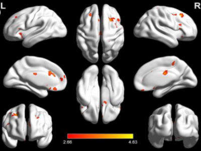
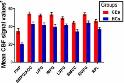
There are no differences in age, gender, and education level between CE and HC (P > 0.05). Cortical regions with significant CBF difference between CE patients and HC were demonstrated in Fig. 1. As it was shown, CE patients had significantly increased CBF compared to controls in the right parahippocampal region, bilateral medial FG/ACC, bilateral IFG, left SFG, bilateral MCC, right MFG (BA8), and right paracentral lobule. Specific comparison of CBF values between those regions were shown in Fig. 2.Discussions and Conclusions
1. The present study demonstrates the hypothesis that CE involves the dysfunction of the visual pathway (i.e. the supplementary eye field located in the MFG, ACC, and frontal eye field (BA8)). 2. Interestingly, the most significant CBF increase occurs in the right parahippocampal region, suggesting the involvement in potential cognitive and mood compensation in CE.4Acknowledgements
No acknowledgement found.References
1. Chen X, Fu Z, Yu J, et al. Prevalence of amblyopia and strabismus in Eastern China: results from screening of preschool children aged 36–72 months British Journal of Ophthalmology 2016;100:515-519.2. Yan, X., Lin, X., Wang, Q., Zhang, Y., Chen, Y., Song, S., & Jiang, T. (2010). Dorsal Visual Pathway Changes in Patients with Comitant Extropia. PLoS ONE, 5(6), e10931. 3. Ze Wang, Geoffrey Aguirre, Hengyi Rao, JiongJiong Wang, Anna R. Childress, John A. Detre, Empirical ASL data analysis using an ASL data processing toolbox: ASLtbx, Magnetic Resonance Imaging, 2008, 26(2):261-9.4. McBain, H. B., MacKenzie, K. A., Hancox, J., Ezra, D. G., Adams, G. G. W., & Newman, S. P. (2016). Does strabismus surgery improve quality and mood, and what factors influence this? Eye, 30(5), 656–667.Figures

Fig 1. Regions of significant CBF increases in CE relative to healthy controls (P < 0.05, GRF corrected). L: left; R: right.

Fig 2. Specific comparison of CBF values between CE and healthy controls in right parahippocampal region, bilateral medial FG/ACC, bilateral IFG, left SFG, bilateral MCC, right MFG (BA8), and right paracentral lobule.