1970
MAGNETIC RESONANCE IMAGING TEXTURE ANALYSIS (MRTA) OF NASOPHARYNGEAL CARCINOMA IN T2W AND CE-T1W IMAGES1RADIOLOGY, THE FIRST AFFLIATED HOSPITAL OF DALIAN MEDICAL UNIVERSITY, DALIAN, China
Synopsis
Nasopharyngeal carcinoma is a common malignant tumour in Asian countries with nearly 80% of them being squamous cell carcinoma. The aim is to investigate the potential of MRI (T2W & CE-T1W) texture analysis to predict response in patients with advanced Nasopharyngeal carcinoma(squamous cell carcinoma).The patients were grouped into Residual/Non-Responders and Non-Residual/Responders based on the post-treatment MR images. Texture analysis was used to find significant parameters. On T2WI, significance were recorded with 2 parameters which showed potential to predict the response to treatment and can be further used in the future studies to predict and alter the treatment course and cycles
BACKGROUND AND PURPOSE
Nasopharyngeal carcinoma is a common malignant tumour in Asian countries[1] of which nearly 80% of them are squamous cell carcinoma. The standard treatment for the patients with NPC is radiotherapy. The survival rates have improved with the introduction of chemo-radiotherapy[2].Patients with advanced NPC(stage III-IVB) generally have poorer prognosis due to treatment failure. The main factors for treatment failure is local recurrence and distant metastasis[3].The pretreatment prediction of response to treatment will help to change the course and aggressiveness of the treatment.
This study is to investigate the potential of MRI (T2W & CET1W) texture analysis to predict response in patients with advanced Nasopharyngeal carcinoma(squamous cell carcinoma).
Materials & methods
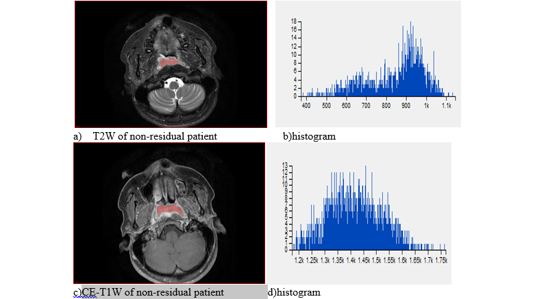
53 patients from 2011-02 to 2017-07 with histologically proven advanced squamous cell carcinoma of nasopharynx were enrolled for this study. They were grouped into 2: Residual/Non-Responders (N=27) & Non-Residual/Responders (n=26) based on the post-treatment/follow up MR images. A total of 53 features were extracted from T2W (n=53) and CE-T1W (N=53) from each patient. Contrasted T1WI, T2WI MRI Digital Imaging and communications in medicine (DICOM) images performed with 1.5 Tesla machine, before treatment, were selected. The TA was performed with Omni-Kinetics Software (GE Healthcare, China);under the guidance of well-experienced mentor, the 3D ROI were delimited manually(freehand), on the axial slice showing the largest tumor section, avoiding large edema, lymph nodes and muscle infiltration. First and second orders texture parameters were selected for statistical analysis on SPSS.24 software; Independent T-test and Mann-Whitney test were used to compare between the two groups; p﹤0.05 symbolized statistical significance. Receiver operating characteristics (ROC) were used to assess sensitivity and specificity.Results
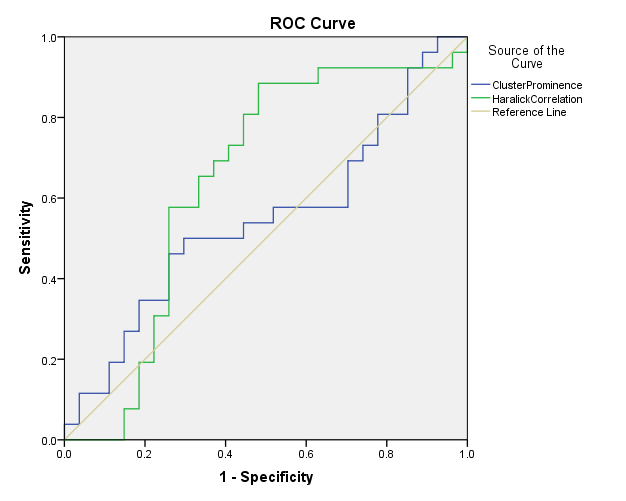
41 patients were male with mean age of 53(+/-10.12) & 12 female patients with mean age of 57(+/-15.38). On T2WI images, we recorded significance with parameters Cluster Prominence (p=0.004, AUC=0.729, sensitivity=67%, specificity=77%) & Haralick Correlation(p=0.049, AUC=0.659, sensitivity=67% specificity=73%).Other parameters in T2W images did not show significance(p>0.05).no significant parameters were obtained from CET1W images (p>0.05).Discussion
In this study, a 2 feature based score was obtained and validated to be an independent predictor of response/progression of advanced NPC. The score was obtained from T2W images alone and had better prognostic value than CET1W images. The present TNM staging based on gross anatomy has a disadvantage that it does'nt take into account the heterogeneity of the tumor lesion. The intra-tumor heterogeneity is found to have crucial clinical significance in diagnosis, staging and prognosis of the disease[4]. Texture analysis is a new and promising research area in the field of medical imaging with potential to diagnose, predict more information about the images obtained after a seemingly routine MRI scan. MRI is routinely used to diagnose and follow up NPC. In contrast to CT , MRI provides better tissue contrast and lesser artifacts. So we obtained features from multi-parametric MR images with T2W & CET1W images. It was found that the model derived from T2W I images faired better than the model got from CET1W images. To develop the model,53 parameters were used which was reduced to set of only 2 potential prognostic parameters/factors. The 2 most important parameters showed significant difference between the Residual/Non-Responders and Non-Residual/Responders. The limitation of this study was that the (i)only the slice with largest tumor size was used for texture analysis (ii) the small sample size obtained after the exclusion criteria. 53 patients could be included from the set of 113 NPC patients.Conclusion
1.The 2 parameters extracted after the texture analysis of T2W images showed potential to predict the response to treatment in patients with NPC. These can be further used in the future studies to predict the prodnosis and thereby alter the treatment course and number of cycles of chemo-radiotherapy.
2.T2W images were better suitable to predict the treatment response than CET1W images.
Acknowledgements
No acknowledgement found.References
1. Chen L, Hu CS, Chen XZ, Hu GQ, Cheng ZB, Sun Y, Li WX, Chen YY, Xie FY, Liang SB, Chen Y, Xu TT, Li B, et al. Concurrent chemoradiotherapy plus adjuvant chemotherapy versus concurrent chemoradiotherapy alone in patients with locoregionally advanced nasopharyngeal carcinoma: a phase 3 multicentre randomised controlled trial. Lancet Oncol. 2012;13:163–171. [PubMed]
2. Saleh-Ebrahimi L, Zwicker F, Muenter MW, Bischof M, Lindel K, Debus J, Huber PE, Roeder F. Intensity modulated radiotherapy (IMRT) combined with concurrent but not adjuvant chemotherapy in primary nasopharyngeal cancer - a retrospective single center analysis. Radiat Oncol. 2013;8:20. [PMC free article] [PubMed]
3. Hou X, Zhao C, Guo Y, Han F, Lu LX, Wu SX, Li S, Huang PY, Huang H, Zhang L. Different clinical significance of pre- and post- treatment plasma Epstein-Barr virus DNA load in nasopharyngeal carcinoma treated with radiotherapy.Clin Oncol (R Coll Radiol) 2011;23:128–133. [PubMed] 4. Sala E, Mema E, Himoto Y, Veeraraghavan H, Brenton JD, Snyder A, Weigelt B, Vargas HA. Unravelling tumour heterogeneity using next-generation imaging: radiomics, radiogenomics, and habitat imaging. Clin Radiol. 2017;72:3–10. [PMC free article] [PubMed]
Figures