1968
One-step high-resolution diffusion weighted imaging in ocular masses and optic nerve using a dedicated surface coil1Radiology Department, Beijing Tongren Hospital, Capital Medical University, Beijing, China, 2Philips Healthcare China, Beijing, China
Synopsis
It is challenging for the routine clinical ocular MRI protocol to use a large FOV covering the whole orbits and sellar region with high spatial resolution relatively. The aim of this study was to evaluate custom-made ocular surface coil in diagnosing images for ocular masses and the optic nerve by comparing TSE DWI images. The dedicated ocular coil obtained large FOV and high spatial resolution images with higher SNR in TSE DW images as examples. The custom-made surface coil can demonstrate ocular masses and the optic nerve more clearly, and provide more details with high SNR in one-step.
Synopsis
It is challenging for the routine clinical ocular MRI protocol to use a large FOV covering the whole orbits and sellar region with high spatial resolution relatively. The aim of this study was to evaluate custom-made ocular surface coil in diagnosing images for ocular masses and the optic nerve by comparing TSE DWI images. The dedicated ocular coil obtained large FOV and high spatial resolution images with higher SNR in TSE DW images as examples. The custom-made surface coil can demonstrate ocular masses and the optic nerve more clearly, and provide more details with high SNR in one-step. Purpose
Routine clinical ocular MRI FOV includes eye globes,
optic nerve and even sellar region, because the ocular masses including retinoblastoma
may invade the optic nerve[1]. The conventional MRI with head coils and
large FOV covers the whole orbits and sellar region. However, the small size
makes it challenging to characterize the ocular masses
and the optic nerve. Besides, it is hard to analyze images acquired with the out-of-factory
head coils, because of the distortion in MRI signal due to adjacent air-filled
bony structures[2,3].
Echo-planar imaging
(EPI) based DWI can help characterize indeterminate orbital masses[4].However, image
with EPI based DWI for ocular masses is challenging due to image distortions
from susceptibility artifacts, making it difficult to evaluate image qualities.
Hence turbo spin-echo (TSE) based DWI was used in this application. On the
other side, SNR is usually lower in TSE DWI signal than EPI DWI; and we
investigated if a dedicated coil could improve the coil sensitivity.
It was proposed small ocular surface coils applied
in high field MRI systems would obtain thinner slice thickness and higher
spatial resolution images with high signal noise ratio relatively. Thereby, to
achieve high spatial resolution high SNR images, the ocular masses imaging studies
would need to use head coils and ocular surface coil successively. Hence
custom-made ocular surface coil was designed to image ocular masses and the optic
nerve together. The aim of this study was to evaluate of TSE DWI using this custom-made
ocular surface coil in diagnosing images for ocular masses and the optic nerve.Purpose
Material and Methods
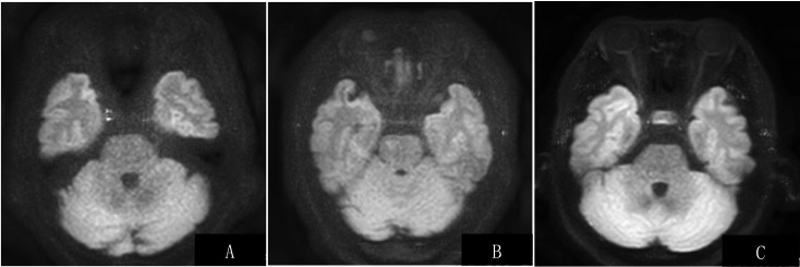
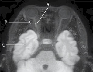
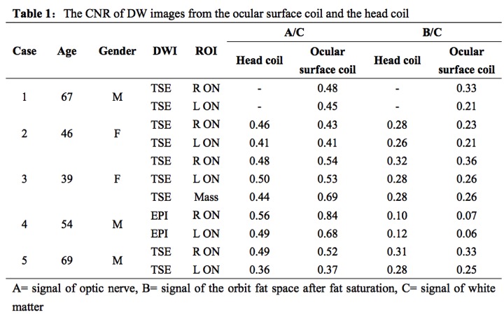
The institutional review board approved the prospective study on five patients(3 men, 2 women; age range 39-69 years)with suspected ocular mass or optical nerve lesions underwent MRI examinations of orbits on a 3.0T MR station (Ingenia 3T, Philips Healthcare, Best, the Netherlands) successively using a 8-channel head coil and a custom-made 360 degree ocular surface coil (Suzhou Medcoil Healthcare Co., Ltd). The ocular coil was tailored to obtain the signal in the deep orbits and sellar region. All patients underwent conventional MRI and turbo spin-echo DWI with b-values of 0 and 900 s/mm with the same sequence parameters. The image quality in DWI was qualitatively graded on by 2 readers in consensus on a 3-point scale from 1 (loss or indistinct), 2(partial and blurred) to 3 (full and clear) for the presence of optic nerve (Figure 1). The contrast-to-noise ratio (CNR) of images from the ocular surface coil was compared with the ones from the head coil. (Figure 2)Results
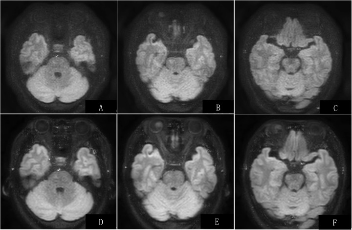
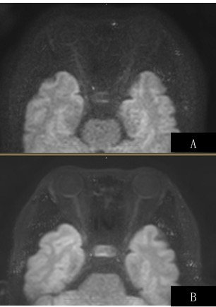
Both ocular masses and optic nerve showed more clearly in the DW images from the ocular surface coil (Figure 3E, 4A) than in the ones from the head coil (Figure 3B, 4B). The DW images from the ocular surface coil dedicated more internal details of the ocular mass than the ones from the head coil (Figure 3B). The presence of optic nerves was totally rated 3 in five patients in the DW images from the ocular surface coil, and rated 1 in two patients, 2 in two patients, and 3 in one patients in the DW images from the head coil. The contrast-to-noise ratio (CNR) of DW images from the ocular surface coil was higher than the ones from the head coil (Table 1).Discussion and Conclusion
This study demonstrated that, comparing to previously used head coil and surface coils, the custom-made dedicated small ocular surface coils generated better images for both ocular masses and the optic nerve simultaneously with high spatial resolution and high SNR. The ocular masses imaging could benefit from its convenience in covering more regions. Both ocular masses and optic nerve showed more clearly and more details in the DW images from the ocular coil than in the images from the head coil. Moreover, the high quality of DW images offers unique insights into the internal microstructure and texture information, particularly with regard to pathology and genomics. It can improve our future ability to study ocular masses and optic nerve pathologies noninvasively, offer information in the differential diagnosis of ocular masses and monitor the changes of their treatment[5,6].Acknowledgements
No acknowledgement found.References
1. de Graaf P, Pouwels PJ, Rodjan F, et al. Single-shot turbo spin-echo diffusion-weighted imaging for retinoblastoma: initial experience. Am J Neuroradiol. 2012;33(1):110-118.
2. Hoch MJ, Bruno MT, Shepherd TM. Advanced MRI of the Optic Nerve. J Neuroophthalmol. 2017;37(2):187-196.
3.Erb-Eigner K, Willerding G, Taupitz M, et al. Diffusion-weighted imaging of ocular melanoma. Invest Radiol. 2013;48(10):702-707.
4. Sepahdari AR, Aakalu VK, Setabutr P, et al. Indeterminate orbital masses: restricted diffusion at MR imaging with echo-planar diffusion-weighted imaging predicts malignancy. Radiology. 2010;256(2):554-564.
5. Sepahdari AR, Kapur R, Aakalu VK, et al. Diffusion-weighted imaging of malignant ocular masses: initial results and directions for further study. Am J Neuroradiol. 2012; 33(2):314-319.
6. Foti PV, Longo A, Reibaldi M, et al. Uveal melanoma: quantitative evaluation of diffusion-weighted MR imaging in the response assessment after proton-beam therapy, long-term follow-up. Radiol Med. 2017;122(2):131-139.
Figures