1963
MRI Assessment of SPION Contrast in the Inner Ear1Physical Sciences Platform, Sunnybrook Research Institute, Toronto, ON, Canada, 2Department of Otolaryngology Head & Neck Surgery, University of Toronto, Toronto, ON, Canada, 3Biological Sciences Platform, Sunnybrook Research Institute, Toronto, ON, Canada, 4Medical Biophysics, University of Toronto, Toronto, ON, Canada, 5Neurosurgery and Pediatric Neurosurgery, Medical University of Lublin, Lublin, Poland
Synopsis
A novel approach to diagnostic imaging and treatment of the inner ear disorders is magnetic targeting of therapy using superparamagnetic iron oxide nanoparticles (SPIONs). SPIONs were deposited onto the round window niche using a surgical approach, and then magnetic targeting was used, in the treatment group, to “pull” the SPIONs further into the inner ear. High resolution T2 weighted imaging was used to assess the treatment. Signal loss was observed in the vestibule and cochlea in both groups, while increased signal loss was observed at the apex of the cochlea in treated animals relative to the control group.
Introduction
Inner ear disorders can cause loss of hearing, vertigo, tinnitus, and loss of balance1. These disorders are difficult to diagnose and treat as the inner ear is housed in dense bone, and further subdivided into different fluid-filled compartments.
There is currently no reliable technique to deliver contrast or therapy to the inner ear for diagnostic imaging or treatment. One approach is to rely on passive diffusion of intratympanic medications through the round window, while the others utilize more invasive techniques such as cochleostomy which can lead to further hearing loss or balance dysfunction2.
A novel approach to diagnostic imaging and treatment of the inner ear disorders is magnetic targeting of therapy using superparamagnetic iron oxide nanoparticles (SPIONs) and an external magnetic field to ‘pull’ the SPIONs deeper into the inner ear3. SPIONs have the additional advantage of being visible on MRI, permitting non-invasive assessment of their distribution, which has not previously been done.
Methods
250 g male Long Evans rats were injected with investigative SPIONs by surgical delivery into the left middle ear. Surgery consisted of a post-auricular approach to the bulla, and subsequent deposit of SPIONs onto the round window niche. The animals were then either imaged immediately (2 control rats) or subjected to magnetic targeting in order to “pull” the SPIONs into the inner ear via the round window (2 experimental rats).
Imaging was performed using a 7T Bruker BioSpec MRI. A 3D RARE sequence, with TR 1800ms, echo train of 40, and spacing of 7.2ms for an effective echo time of 100.8ms, 32x32x6.4mm3 field of view, and a resolution of 0.1mm3 in order to visualize the structures of the inner ear.
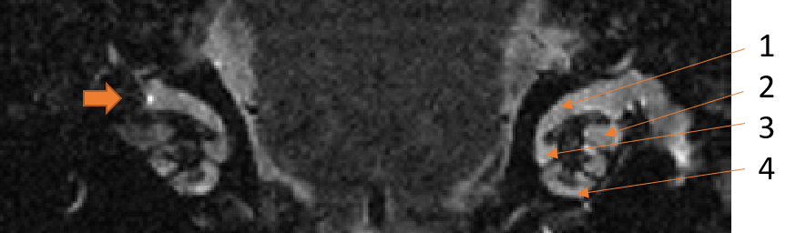
Regions of interest were drawn on coronal sections, at 4 different ‘levels’ of the cochlea. The signal intensity of the treated ear was calculated for each level relative to the signal intensity at the same location in the untreated ear.
Hearing was evaluated using Auditory Brainstem Response (ABR). ABR was performed before, 1 week, and 6 weeks after magnetic delivery to confirm normal hearing.
Results
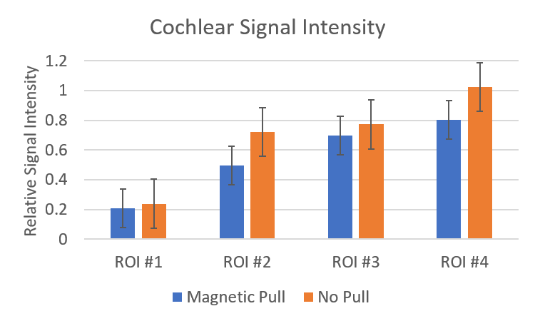
Decreased signal intensity was observed in the treated cochlea of all experimental rats compared to control rats (Figure 1). The greatest signal decrease was seen nearest to the site of the injection, with a smaller signal decrease deeper into the cochlea (Figure 2). In the experimental rats treated with magnetic targeting, there was a further small signal decrease at the apex of the cochlea (0.80±0.15) which was not observed in the control group without magnetic targeting.
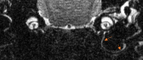
A significant signal decrease in the vestibule was also observed in all animals on the treated side only (Figure 3).
Discusson & Conclusions
The signal decrease in both the cochlea and the vestibule following SPION injection and magnetic targeting indicates that this treatment is reaching both of these structures. The additional signal decrease observed in the experimental animals suggests that the magnetic targeting is improving the penetration of the SPION treatment into the inner ear.Acknowledgements
No acknowledgement found.References
1. E. Smouha. Inner ear disorders. Review. NeuroRehabilitation. 2013;32(3):455-462.
2. Zou J, Zhang W, Poe D, et al. MRI manifestation of novel superparamagnetic iron oxide nanoparticles in the rat inner ear. Nanomedicine. 2010;5(5):739-754.
3. Sarwar A, Nemirovski A, Shapiro B. Optimal Halbach Permanent Magnet Designs for Maximally Pulling and Pushing Nanoparticles. J Magn Magn Mater. 2012;324(5):742-754.
Figures