1961
Compressed-Sensing Accelerated 3-Dimensional Magnetic Resonance Imaging of Inner Ear: A Feasibility Study of Volunteer1Radiology, Peking University First Hospital, Beijing, China, 2Philips Healthcare, Beijing, China
Synopsis
Compressed-Sensing (CS) accelerated 3-dimensional magnetic resonance imaging (MRI) does not reduce image quality even with higher image quality scores compared to conventional MRI of inner ear, while significantly shortening the imaging time. It is a feasible protocol in inner ear imaging.
Introduction
Compressed-Sensing (CS) algorithm is a newer strategy to accelerate data acquisition in 3-dimensional (3D) MRI. Some studies have reported the clinical feasibility of 3D-MR angiography (MRA) and 3D-MR cholangiopancreatography (MRCP) using CS algorithm 1-3. Our purpose is to evaluate image quality of CS accelerated 3D-MRI of inner ear.Methods
From June 2017 to July 2017, twenty volunteers (11 Men and 9 Women) ranging in age from 24 to 31 years (mean 26 years) were included in this study. They were examined utilizing both CS accelerated 3D-MRI (CS-MRI) and conventional 3D-MRI (Con-MRI) of inner ear on 3T MR system. Multiplanar reconstruction (MPR) images perpendicular to the long axis of the internal auditory canal (IAC) were reconstructed to observe the four nerves (facial nerve, cochlear nerve, and inferior and superior vestibular nerve) within the IAC. The image quality of both sides of facial nerve, cochlear nerve, MPR images of IAC, cochlear turn, three semicircular canals and trigeminus nerve were assessed by two radiologists (2 and 6 years of experience in MR imaging), respectively. Scores of 1, 2, 3, 4 and 5 were assigned to non-diagnostic, poor, fair, good, and excellent image quality. Inter-observer agreement was evaluated by Kappa analysis. Nonparametric test (Wilcoxon test) was performed to compare the image quality scores between CS-MRI and Con-MRI.Results
1. The inter-observer agreement analyses showed good agreements regarding image quality assessment in CS-MRI (Kappa=1, P<0.001) and Con-MRI (Kappa=0.876, P<0.001).
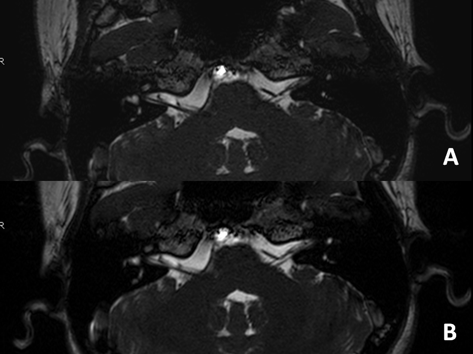
2. The image quality scores of right facial nerve assessed by radiologist with 2 years experience gained significantly higher in CS-MRI compared to Con-MRI (4.95±0.22 for CS-MRI, 4.55±0.76 for Con-MRI, Z = -2.07, P = 0.038). The image quality scores of right facial nerve, right cochlear nerve, right MPR images of IAC, and right three semicircular canals assessed by radiologist with 6 years experience gained significantly higher in CS-MRI compared to Con-MRI (P<0.05) (Table 1, Fig. 1). No significant difference of other scores in CS-MRI compared to Con-MRI (P>0.05).
Discussion
Our results show that, CS-MRI does not reduce image quality compared to Con-MRI of inner ear. Even some image quality scores of CS-MRI showed significantly higher than that of Con-MRI. In our study, the mean acquisition time of CS-MRI in our study was about 3 min, whereas Con-MRI took about 6 min. Scanning time was nearly 50% saved. So we think higher image quality score of CS-MRI mainly because of the less movement from the patients in shorter scanning time. So CS-MRI is a feasible protocol in inner ear imaging. However, further studies included patients of inner ear should be done to test the clinical feasibility of CS accelerated 3D-MRI.Conclusion
CS accelerated 3D-MRI is a feasible protocol in inner ear imaging. Future protocol design could use CS to reduce the scanning time.Acknowledgements
No acknowledgement found.References
[1] Li B, Li H, Dong L, Huang G. Fast carotid artery MR angiography with compressed sensing based three-dimensional time-of-flight sequence. Magn Reson Imaging. 2017; 43:129-135.
[2] Fushimi Y, Fujimoto K, Okada T, ea al. Compressed Sensing 3-Dimensional Time-of-Flight Magnetic Resonance Angiography for Cerebral Aneurysms: Optimization and Evaluation. Invest Radiol. 2016; 51(4):228-235.
[3] Zhu L, Wu X, Sun Z, et al. Compressed-Sensing Accelerated 3-Dimensional Magnetic Resonance Cholangiopancreatography: Application in Suspected Pancreatic Diseases. Invest Radiol. Sep 26, 2017.
Figures