1944
Altered marginal division connectivity in Parkinson disease with mild cognitive impairment revealed by resting-state fMRILi mingge1,2, Chen yuanyuan3, Feng jie1, Zhang shiyu1, Lou xin1, and Ma lin1
1Chinese PLA general hospital, beijing, China, 2Nankai University, tianjin, China, 3Tianjin University, tianjin, China
Synopsis
The marginal division (MrD) functional connectivity is disrupted during mild cognitive impairment in Parkinson’s disease.
Objective
The marginal division (MrD) of neostriatum is a neostriatum subregion between the putamen and the globus pallidus, and it is an important subcortical center that is involved in learning and memory. In this study, we used resting state-functional magnetic resonance imaging (rs-fMRI) to explore altered MrD functional connectivity (FC) in Parkinson’s disease patients with mild cognitive impairment.Materials and methods
28 PD patients with mild cognitive impairment (PD-MCI) (17 males,11 females,mean age 64.75±8.17 years) and 25 PD patients having no cognitive impairment (PD-NCI) (9 males,16 females,mean age 59.20±8.56 years) recruited in the cross-sectional study from Feburary 2017 to Ocotuber 2017. Meanwhile 29 healthy age-matched controls (11 males,18 females,mean age 61.90±7.05 years) were included. MRI exams including T2 weighted image, 3-dimensional fast spoiled gradient recalled echo (3D FSPGR), and rs-fMRI were performed in all participants. All subjects were enderwent neurological and cognitive test ,including Unified Parkinson’s Disease Rating Scale (UPDRS), Mini Mental State Examination (MMSE), Beijing Version of Montreal Cognitive Assessment (MoCA), Clinical Dementia Rating (CDR) before MRI scan. MrD was defined using manual delineation on structural images, and was selected as the seed to calculate the FC. All MR structural and functional images were processed using Statistical Parametric Mapping 12 (SPM12) and the data processing assistant for resting-state fMRI (DPARSF). The clinical data including the age, BMI, education, disease duration, MMSE score, MoCA score, UPDRS III score and H-Y stage, were analyzed by one-way analysis of variance (ANOVA) test, chi-square test, two-sample t-test. Significant difference was set at a p value of < 0.05. The statistical analysis was performed using SPSS 17.0. Two-sample t-tests were performed to detect differences in FC of MrD among the PD-MCI, PD-NCI and healthy control groups. Significance was set at a p value of <0.001 without correction. The minimal number of contiguous voxels was set at 10. The statistical analysis was performed by SPM 12 software.Results
The three subject groups showed differences in age (p=0.046), MMSE score (p=0.000), MoCA score (p=0.000), CDR scores (p=0.000), but no diference in gender (p=0.667), BMI (p=1.000), education (p=0.253). The disease duration (p=0.072) and H-Y stage (p=0.051) showed no differences between PD-MCI and PD-NCI patients groups, but he UPDRS III score (p=0.01) showed difference. Compared with healthy controls, a different decreased FC pattern was observed between the MrD and other brain regions. We also found increased FC between the left MrD and the right MrD in PD patients.Compared with PD-NCI, the PD-MCI patients showed dcreased MrD FC with insula, inferior frontal gyrus, putamen and middle occipital gyrus. Increased MrD FC with middle cingulum, precuneus, supplement motor area, paracentral lobe, and inferior frontal gyrus.Conclusion
Our findings demonstrate that the MrD functional connectivity is disrupted during mild cognitive impairment in Parkinson’s disease. However, the correlation between MrD functional connectivity strength with the cognitive abilities scores in PD patients requires further investigation.Acknowledgements
No acknowledgement found.References
No reference found.Figures
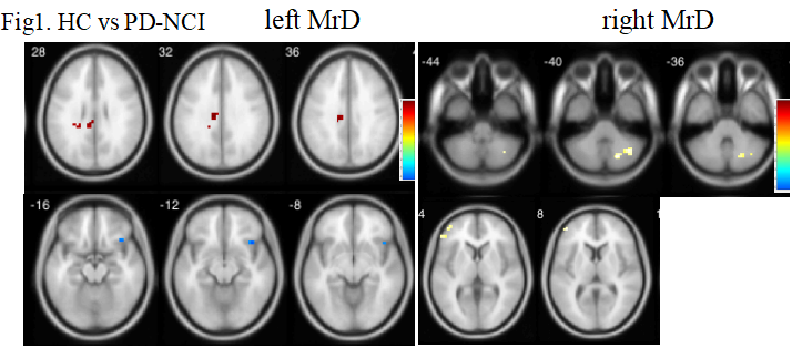
Fig1. Comparison of MrD FC in healthy control subjects and PD-NCI patients.Note: HC, healthy control; PD-NCI, Parkinson’s disease patients having no cognitive impairment.
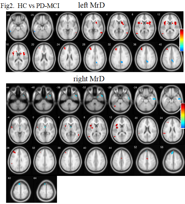
Fig2. Comparison of MrD FC in healthy control subjects and PD-MCI patients. Note: HC, healthy control; PD-MCI, Parkinson’s disease patients with mild cognitive impairment.
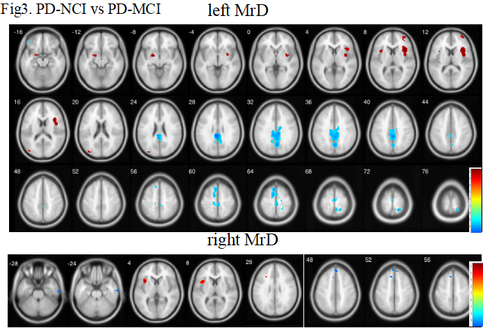
Fig3. Comparison of MrD FC in PD-NCI patients and PD-MCI patients. Note: PD-NCI, Parkinson’s disease patients having no cognitive impairment; PD-MCI, Parkinson’s disease patients with mild cognitive impairment.