1933
Evaluating the sensitivity of univariate and multivariate techniques on diffusion-derived metrics in classification of early Parkinson’s disease patients1Imaging, Cleveland Clinic Lou Ruvo Center for Brain Health, Las Vegas, NV, United States
Synopsis
In this study, we utilized the diffusion MRI (
Introduction
The pathologic development of Parkinson’s disease (PD) appears to be related to the spread of abnormal synuclein in a largely caudal-rostral direction in the CNS1–3. Diffusion tensor imaging (DTI) is capable of delineating in vivo microstructural changes of white matter tracts noninvasively. DTI can detect alterations in white matter in early stages of PD4, with potential to develop into important biomarker in understanding PD. However, studies utilizing either voxelwise comparisons or skeleton-wise comparisons have produced conflicting conclusions in diffusion derived measures of the same region4. Moreover, most of the studies with early PD suffer from low sample size resulting in low statistical power which may also contribute to conflicting results. Hence, in this study, we utilized multi-site DTI data from a large cohort of healthy controls (HC) and early PD-patients from the Parkinson’s Progressive Markers Initiative (PPMI) database5, and performed both voxelwise and skeleton-wise comparisons to classify PD from HC. Both tract-based spatial statistics (TBSS), and tensor based registration (DTI-TK)6 algorithm were used for skeleton-wise comparisons. We also extracted diffusion-derived measures in the 20 major white matter (WM) tracts of JHU atlas7 and evaluated their sensitivity and specificity in the classification of PD from HC. Furthermore, both univariate and multivariate techniques were used to investigate the strengths and weaknesses of each technique.Methods
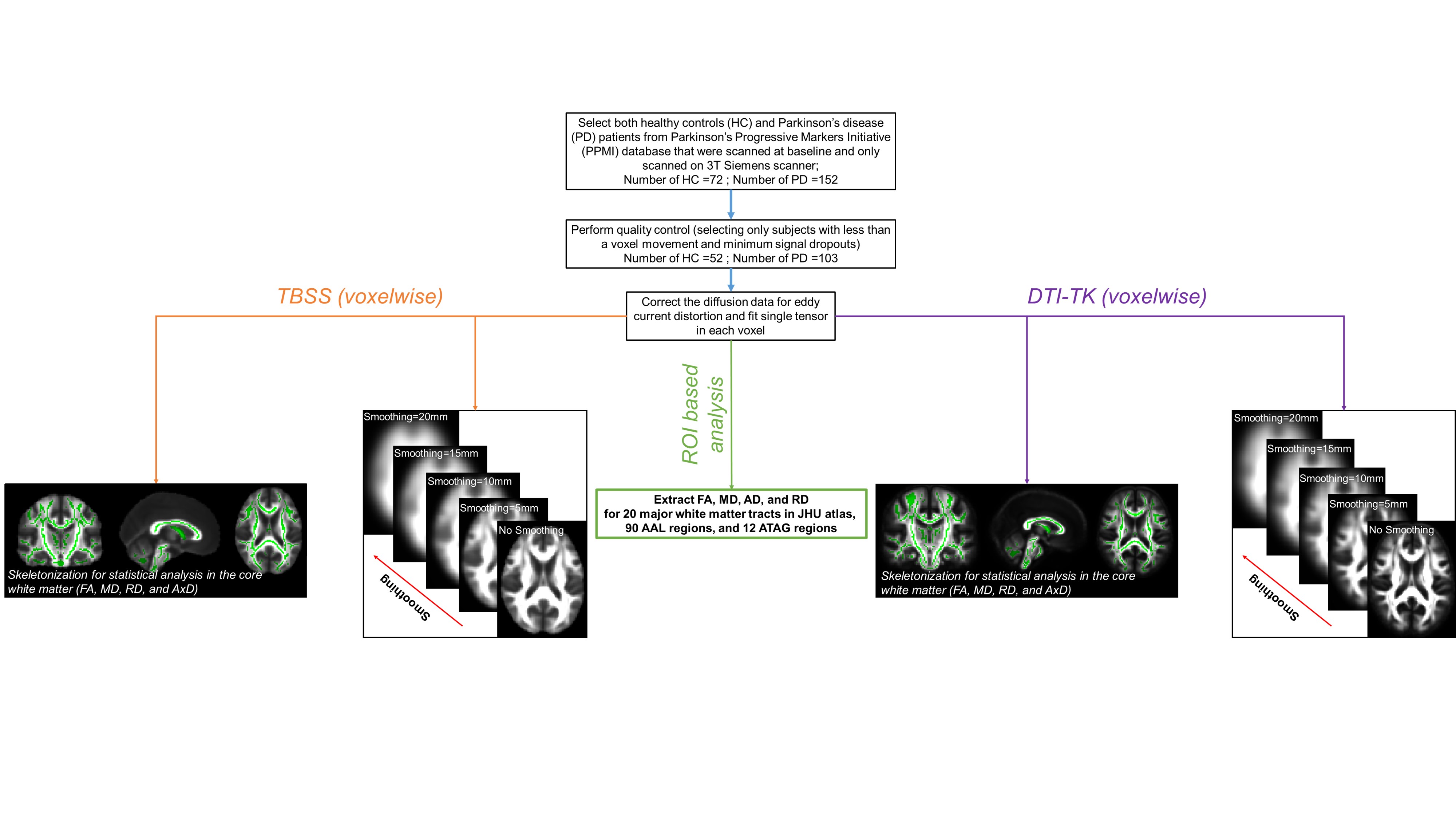
Subjects: Diffusion-MRI (dMRI) data from PPMI database from 72 (24 female) HC and 152 early PD-subjects (54 female) was used in this study. Imaging parameters are described in detail at http://www.ppmi-info.org/5. Only data from 3T Siemens scanners with the first visit were used to ensure uniformity of diffusion data. dMRI dataset of each PD patient were corrected for eddy-current distortions using FSL, and the average motion across diffusion directions was calculated. Only those patients with less than a voxel movement (on an average) were used in this study. This criterion yielded 52 HC (17F, age: 60.69±11.19years, years of education (YOE): 15.58±3.09years) and 103 PD patients (age: 61.11±9.92years, (YOE): 15.36±3.02years, total MDS-UPDRS: 19.5±8.22, disease duration: 10.69±12.79months). Data preprocessing: Single tensor was fitted for every subject using FSL and a voxel-wise estimate of fractional anisotropy (FA), axial diffusivity (AD), radial diffusivity (RD), and mean diffusivity (MD) was calculated. In addition, a tensor map was saved for every subject. Smoothing: Smoothing of the raw tensor files was done from 0mm to 20mm to evaluate the effect of smoothing8 on voxelwise and skeleton-wise measures. Skeleton-wise analysis: Standard TBSS and DTI-TK was used for both groups at each smoothing level. Extraction of dMRI derived measures from JHU atlas: dMRI derived measures were extracted for each subject after DTI-TK registration of each subject to MNI152 space. Voxelwise analysis: In addition, standard voxelwise comparison was performed between each group at each smoothing level. Statistical analysis: The demographics were used as the regressor of no interest and all statistical analysis, both univariate and multivariate measures, at each step was conducted in PALM9. In addition, voxelwise regression analysis was conducted to see the extent of disease severity and disease duration on WM abnormalities, if any, between HC and PD patients.Results
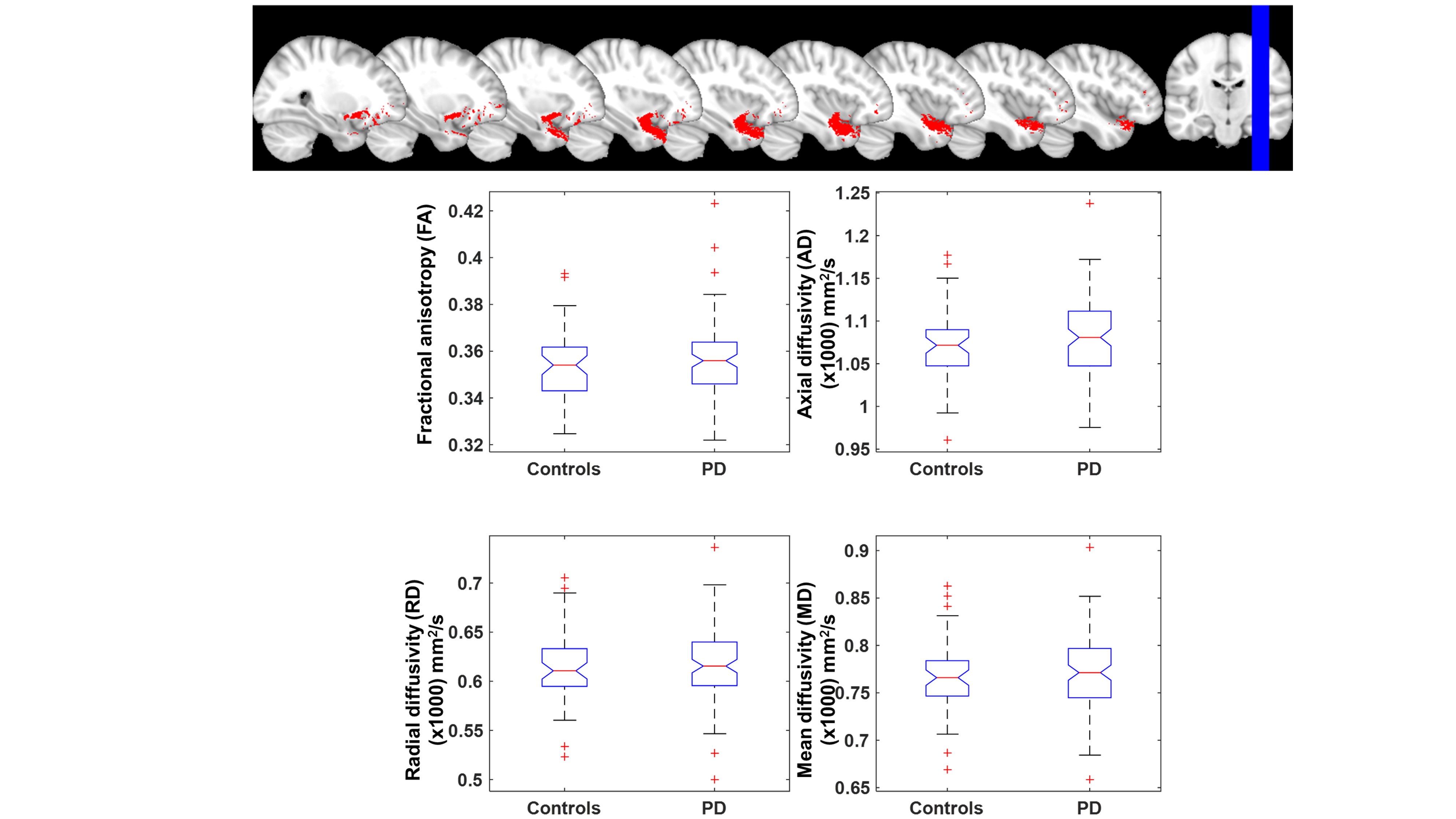
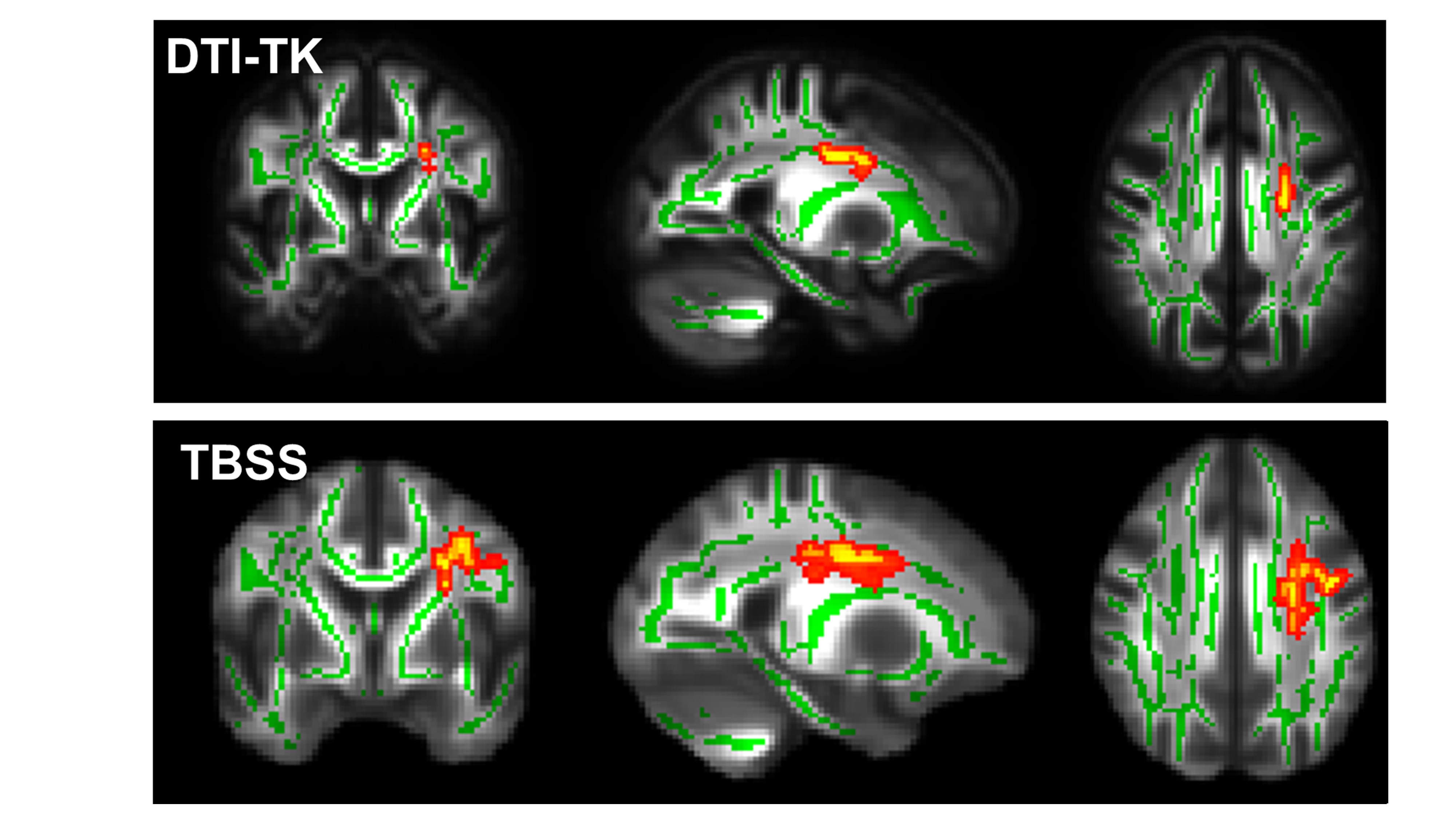
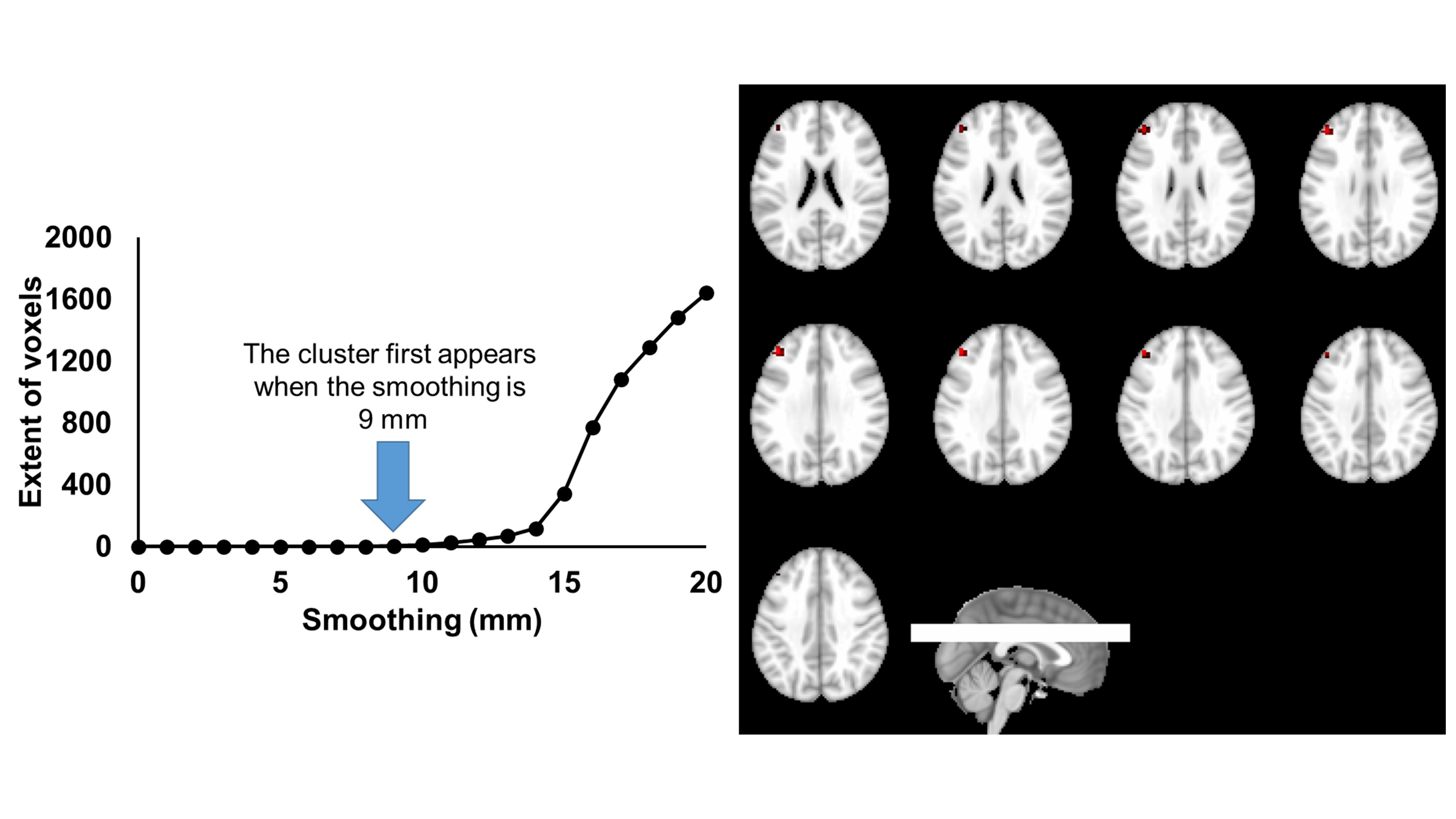
Fig.1 shows a brief flowchart of the entire method used in this study. Both univariate and multivariate analysis of 20 WM tracts revealed no differences between HC and PD patients. However, multivariate analysis revealed uncinate fasciculus in the right hemisphere to be positively associated with disease duration (Fig.2). The skeleton-wise analysis revealed FA of left corticospinal tract to be negatively associated with disease duration in PD patients with no smoothing, and was consistently found regardless of the algorithm used (Fig.3). However, this difference vanished when smoothing was employed. Voxelwise measures revealed a cluster in the right frontal cortex where MD was lower in PD patients, as shown in Fig.4.Discussion and Conclusion:
In this study, we revealed (a) multivariate methods are more powerful for region of interest analysis to study association with disease symptom; (b) Both DTI-TK and TBSS algorithm perform equally well in understanding disease progression and WM abnormalities; and (c) smoothing plays an important role in classification of HC and PD patients. These findings are consistent with the known pathology of PD and may provide the clinicians better understanding of pathophysiology of PD as the disease progresses which is currently poorly understood.Acknowledgements
The study issupported by the National Institutes of Health (grant number 1R01EB014284 and P20GM109025). PPMI is sponsored and partially funded by The Michael J. Fox Foundation for Parkinson’s Research (MJFF). Other funding partners include a consortium of industry players, non-profit organizations and private individuals (for a full list see http://www.ppmi-info.org/about-ppmi/who-we-are/study-sponsors/).References
1 Goedert M, Spillantini MG, Del Tredici K, Braak H. 100 years of Lewy pathology. Nat Rev Neurol 2013; 9: 13–24.
2 Braak H, Del Tredici K, Rub U, de Vos RAI, Jansen Steur ENH, Braak E. Staging of brain pathology related to sporadic Parkinson’s disease. Neurobiol Aging 2003; 24: 197–211.
3 Braak H, Bohl JR, Muller CM, Rub U, de Vos RAI, Del Tredici K. Stanley Fahn Lecture 2005: The staging procedure for the inclusion body pathology associated with sporadic Parkinson’s disease reconsidered. Mov Disord 2006; 21: 2042–51.
4 Hall JM, Ehgoetz Martens KA, Walton CC, et al. Diffusion alterations associated with Parkinson’s disease symptomatology: A review of the literature. Parkinsonism Relat Disord 2016; published online Sept. DOI:10.1016/j.parkreldis.2016.09.026.
5 www.ppmi-info.org..
6 Zhang H, Yushkevich PA, Alexander DC, Gee JC. Deformable registration of diffusion tensor MR images with explicit orientation optimization. Med Image Anal 2006; 10: 764–85.
7 Hua K, Zhang J, Wakana S, et al. Tract probability maps in stereotaxic spaces: analyses of white matter anatomy and tract-specific quantification. Neuroimage 2008; 39: 336–47.
8 Jones DK, Symms MR, Cercignani M, Howard RJ. The effect of filter size on VBM analyses of DT-MRI data. Neuroimage 2005; 26: 546–54.
9 Winkler AM, Ridgway GR, Douaud G, Nichols TE, Smith SM. Faster permutation inference in brain imaging. Neuroimage 2016; 141: 502–16.
Figures