1928
Focal Cortical thickness and Subcortical volume changes differ between Parkinson disease subtypes1Department of Radiology, Affiliated Hospital of GuizhouMedical University, Guiyang, China
Synopsis
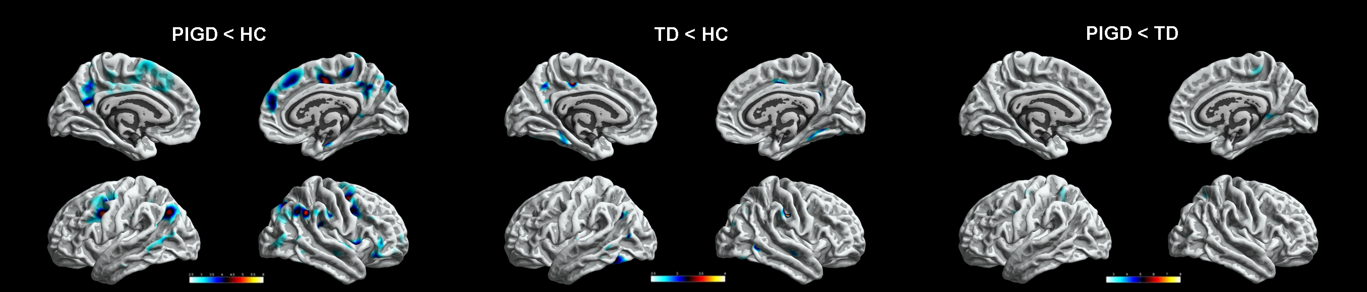
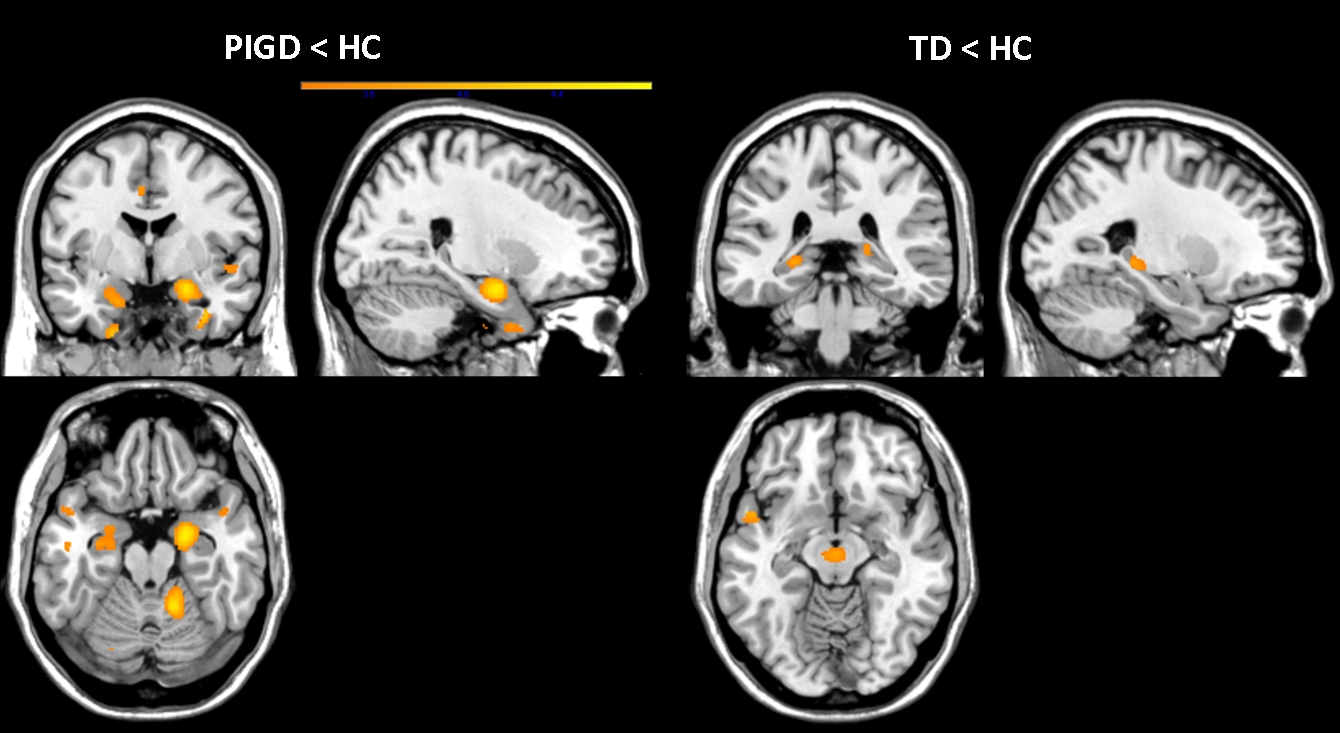
Previous morphometric studies of Parkinson disease (PD) were mainly conducted by measuring gray matter volume and cortical thickness, and little attention has been paid to whether structure MRI improves PD diagnosis or helps differentiating between phenotypes, such as postural instability gait difficulty (PIGD) and tremor dominant (TD). From this study, compared with the control group, PIGD patients had significantly thinning cortical thickness in multiple brain regions, such as bilateral inferiorparietal, paracentral, postiocingulate, superiorfrontal, precuneus, caudalmiddlefrontal, superfrontal and right parsorbitals. TD patients had significantly thinning cortical thickness in left posteriocingulate, inferioparietal and right superiofrontal, superiortemporal, postcentral, precuneus, fusiform and parahippacampal . In addition, subcortical volume atrophy was identified in the bilateral hippocampus and bilateral amygdala of the patients with PIGD, only little bilateral hippocampus changes was found in the TD group.
Introduction
Previous morphometric studies of Parkinson disease (PD) were mainly conducted by measuring gray matter volume and cortical thickness, and little attention has been paid to whether structure MRI improves PD diagnosis or helps differentiating between phenotypes, such as postural instability gait difficulty (PIGD) and tremor dominant (TD).Methods
38 PD patients, including 16 TD subtype, 22 PIGD subtype, and 23 matched healthy control subjects underwent 3.0 Tesla high-resolution structural MRI scanning. Cortical thickness and subcortical volumetric analysis were estimated using an automated Computational Anatomy Toolbox ( CAT12) toolbox.Results
Compared with the control group, PIGD patients had significantly thinning cortical thickness in multiple brain regions, such as bilateral inferiorparietal, paracentral, postiocingulate, superiorfrontal, precuneus, caudalmiddlefrontal, superfrontal and right parsorbitals. TD patients had significantly thinning cortical thickness in left posteriocingulate, inferioparietal and right superiofrontal, superiortemporal, postcentral, precuneus, fusiform and parahippacampal . In addition, subcortical volume atrophy was identified in the bilateral hippocampus and bilateral amygdala of the patients with PIGD, 1 only little bilateral hippocampus changes was found in the TD group.Discsussion
Morphometric abnormalities were greater in the PIGD subtype than in the TD subtype, the disparate patterns of cortical and subcortical degeneration can explain the differences in symptoms between the PD subtypes. Further studies are needed to identify the clinical correlates of the structural abnormalities observed in PD patients.Acknowledgements
This work is supported by grants from the Natural Science Foundation of China (8156070059) and Science and Technology Department of Guizhou Province-Guizhou medical university, Qiankehe LG[2012]024, TN2014-51.References
[1] Front Neurol. 2017 Aug 24;8:428. [2] Mov Disord. 2014 Jan;29(1):122-6. [3] Neuroimage Clin. 2016 Dec 21;13:405-414. [4] CNS Neurosci Ther. 2016 May;22(5):360-7. [5] PLoS One. 2013 May 22;8(5):e64222.. [6] Neurology. 2013 Apr 16;80(16):1476-84.Figures