1926
Quantifying nigral degeneration indicates rapid eye movement sleep behavior disorder being a predictor of Parkinson's disease1Department of Radiology, Osaka University Graduate School of Medicine, Suita, Japan, 2Department of Diagnostic and Interventional Radiology, Osaka University Graduate School of Medicine, Suita, Japan, 3Department of neurology, Osaka University Graduate School of Medicine, Suita, Japan, 4Department of Psychiatry, Osaka University Graduate School of Medicine, Suita, Japan, 5Departments of Biomedical Engineering and Radiology, Cornell University, New York, NY, United States
Synopsis
RBD is thought to be prodromal Parkinson's disease (PD), so we aimed to assess the utility of rapid eye movement sleep behavior disorder (RBD) as a predictor of PD using neuromelanin imaging and quantitative susceptibility mapping (QSM). Our results indicated that RBD-related dopamine cell loss and iron deposition in the substantia nigra pars compacta occur in the developmental process of PD. Thus, we conclude that RBD is prodromal PD and quantifying nigral degeneration in RBD is useful in predicting PD.
PURPOSE:
To assess the utility of rapid eye movement sleep behavior disorder (RBD) as a predictor of Parkinson's disease (PD) using neuromelanin imaging and quantitative susceptibility mapping (QSM) in a quantitative and reproducible manner.METHODS:
Ten patients with RBD (RBD group), 18 patients (PD group) with early PD (Hoehn and Yahr scale: 1-2) and 25 age-matched healthy controls (HC group) underwent neuromelanin imaging and QSM and three-dimensional T1W imaging on a 3T magnetic resonance imager. Both neuromelanin and QSM values of the substantia nigra pars compacta (SNpc) were obtained using a region of interest (ROI) based automated segmentation system with the voxel-based morphometric technique. All neuromelanin and QSM images were coregistered with 3D T1w structural images and were spatially normalized with a 1-mm voxel size using Statistical Parametric Mapping 12 software, thus allowing voxel-based measurement with automatic setting of the multislice ROI totally encompassing the entire SNpc. The spatially normalized images of all subjects were smoothed to remove image noise using an isotropic 4-mm full-width Gaussian kernel. Then, the SNpc ROI was placed on each image. Signal to noise ratio (SNR) of the SNpc in the neuromelanin images was calculated on the basis of mean value of the automatically segmented background region (tegmentum). Neuromelanin area was defined to be the area with an SNR higher than that in the background region. The significance of the intergroup differences was tested for each neuromelanin area and QSM value using analysis of variance (ANOVA) with Tukey's post hoc test, and the correlation with the group was tested for each neuromelanin area and QSM value. The diagnostic performance of each neuromelanin imaging and QSM for RBD and early PD was assessed using receiver operating characteristic (ROC) analysis.RESULTS:
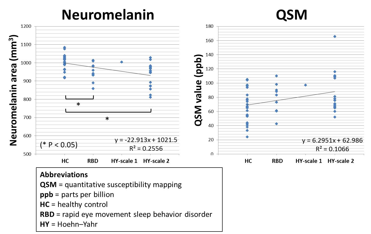
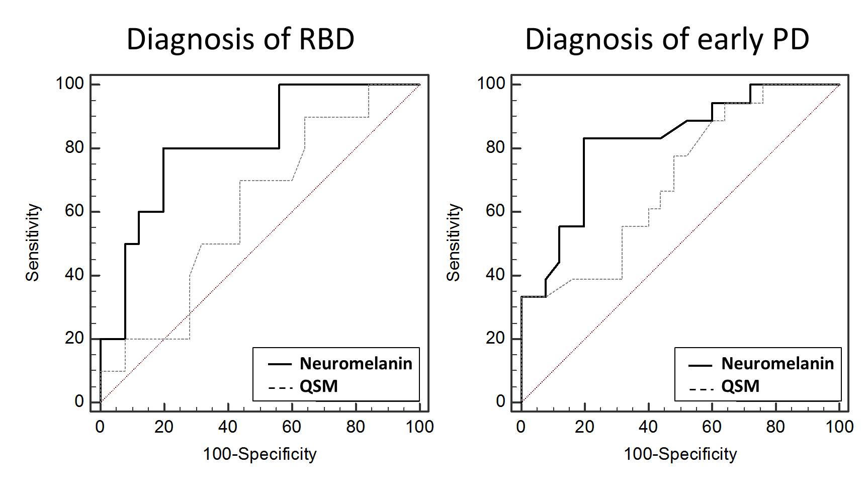
Neuromelanin area was significantly less in both RBD and PD group (RBD: 950.22 ± 49.97 mm3; PD: 936.79 ± 65.80 mm3) than in HC group (1004.69 ± 40.52 mm3) (P < 0.05), by contrast, no significant intergroup difference was shown in the QSM value (RBD: 77.90 ± 20.00 ppb; PD: 87.67 ± 27.90 ppb; HC: 68.46 ± 23.75 ppb). A slight correlation with the group was found in each neuromelanin area (|r| : 0.49) and QSM value(|r| : 0.32) (Fig. 1). The area under the ROC curve (Az [95% confidence interval]) for RBD was 0.81 for neuromelanin area and 0.61 for QSM value. The area under the ROC curve (Az [95% confidence interval]) for early PD was 0.82 for neuromelanin area and 0.70 for QSM value (Fig. 2).DISCUSSION:
RBD is characterized by loss of rapid-eye-movement atonia of sleep and is thought to be a precursor to a synucleinopathy such as PD.1 PD is characterized pathologically by dopaminergic neurodegeneration and iron overload in the SNpc.2, 3 When evaluating the neurodegenerative changes of the SNpc in PD patients, quantifying nigral changes using each neuromelanin imaging and QSM is clinically applicable.4, 5 Neuropathological study suggested that the speed of neurodegeneration is accelerated before reaching a threshold for PD symptoms,6 which indicates the clinical importance of monitoring the early stages in the development of PD. In addition, previous neuromelanin MRI study indicated the clinical usefulness for the diagnosis of early PD stages.7 According to our results, quantifying nigral degeneration using neuromelanin imaging and QSM indicates that RBD may be the PD subtype and initial manifestation preceding PD, thus can provide clinically useful information in the prediction or early diagnosis of PD.CONCLUSION:
RBD-related dopamine cell loss and iron deposition in the SNpc indicated that RBD is prodromal PD and quantifying nigral degeneration in RBD is useful in predicting PD.Acknowledgements
No acknowledgement found.References
1. Chen MC, Yu H, Huang ZL, et al. Rapid eye movement sleep behavior disorder. Curr Opin Neurobiol. 2013;23(5):793-798.
2. GREENFIELD JG, BOSANQUET FD. The brain-stem lesions in Parkinsonism. J Neurol Neurosurg Psychiatry. 1953;16(4):213-226.
3. Graham JM, Paley MN, Grünewald RA, et al. Brain iron deposition in Parkinson's disease imaged using the PRIME magnetic resonance sequence. Brain. 2000;12:2423-2431.
4. Ogisu K, Kudo K, Sasaki M, et al. 3D neuromelanin-sensitive magnetic resonance imaging with semi-automated volume measurement of the substantia nigra pars compacta for diagnosis of Parkinson's disease. Neuroradiology. 2013;55(6):719-724.
5. Azuma M, Hirai T, Yamada K, et al. Lateral Asymmetry and Spatial Difference of Iron Deposition in the Substantia Nigra of Patients with Parkinson Disease Measured with Quantitative Susceptibility Mapping. Am J Neuroradiol. 2016;37:782-788.
6. Lang AE. The progression of Parkinson disease: a hypothesis. Neurology. 2007;68(12):948-952. 7. Ohtsuka C, Sasaki M, Konno K, et al. Changes in substantia nigra and locus coeruleus in patients with early-stage Parkinson's disease using neuromelanin-sensitive MR imaging. Neurosci Lett. 2013;541:93-98.
Figures