1917
The ischemic penumbra assessment using 3D ASL at different post labeling delays in patients with unilateral middle cerebral artery severe stenosis or occlusion1Radiology, The First Affiliated Hospital of Dalian Medical University, Dalian, Dalian, China, 2Radiology, The First Affiliated Hospital of Dalian Medical University, Dalian, China
Synopsis
It is necessary to consider the different PLDs to assess IP by 3D pCASL in ischemic cerebrovascular disease.
Target audience:
Patients with unilateral MCA severe stenosisor occlusion diagnosed by MRA or CTAPurpose
To evaluate ischemic penumbra (IP) using three-dimensional pseudo continuous arterial spin labeling (3D pCASL) at PLD of 1.5 s and 2.5 s in patients with ischemic cerebrovascular diseaseMethods:
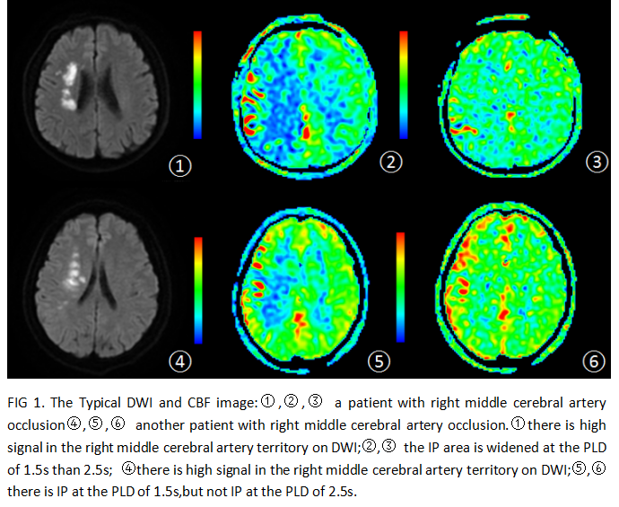
Twenty-six patients (mean age, 60±12 years; 16 men and 10 females) with unilateral middle cerebral artery (MCA) severe stenosis or occlusion were enrolled into the present study, underwent MRI scan especially 3D pCASL with PLDs of 1.5s and 2.5s and DWI. The IP was first observed according to mismatched CBF-DWI region. The mean CBF (CBF1.5 vs. CBF2.5, mL/100g per minute) values and the mean area (mm2) of IP were measured at PLDs of 1.5s and 2.5s. Comparisons of the mean CBF values and the mean IP area between the two PLDs were analyzed using paired T test. Compared with the positive detection rate of IP by Chi-square test.Results:
The detection rate of IP increased at the PLD of 1.5s (21/26, 80.77%) than 2.5s (6/26, 23.08%) (P=0.000). The mean CBF1.5 and CBF2.5 values of IP were 12.32±1.66 vs.18.84±1.44 (P=0.002). The mean IP area was also significantly widened at the PLD of 1.5s (4273.17±611.17) than 2.5s (1074.50±955.32, P=0.01).Conclusions:
IP detection and areas result from different PLD using 3D ASL and DWI in patients with ischemic cerebrovascular disease. The higher detection, decreased CBF and wider region of IP are present at the PLD of 1.5s.Clinical significance:
It is necessary to consider the different PLDs to assess IP by 3D pCASL in ischemic cerebrovascular disease.Acknowledgements
No acknowledgement found.References
[1] Qiu D, Straka M, Zun Z, et al. CBF measurements using multidelay pseudocontinuous and velocity-selective arterial spin labeling in patients with long arterial transit delays: comparison with xenon CT CBF[J]. Magn Reson Imaging,2012,36(1):110–119.
[2] Qiu M, Paul Maguire R, Arora J, et al. Arterial transit time effects in pulsed arterial spin labeling CBF mapping: insight from a PET and MR study in normal human subjects. Magn Reson Med,2010,63(2):374–384.
[3] Bokkers RP, Bremmer JP, van Berckel BN,et al. Arterial spin labeling perfusion MRI at multiple delay times: a correlative study with H (2)(15)O positron emission tomography in patients with symptomatic carotid artery occlusion[J]. Cereb Blood Flow Metab,2010,30(1):222–229.
[4] Campbell AM, Beaulieu C. Pulsed arterial spin labeling parameter optimizationfor an elderly population[J].Magn Reson Imaging 2006,23(3):398–403 .
Figures