1914
Comparison of Image Reconstruction Algorithms of “Flexible PET/MRI” with and without Non-Local Mean Regularization.1Department of Diagnostic Imaging and Nuclear Medicine, Kyoto University Graduate School of Medicine, Kyoto, Japan, 2Human Brain Research Center, Kyoto University Graduate School of Medicine, Kyoto, Japan
Synopsis
Flexible PET (fxPET) is a prototype of MR-compatible mobile PET system. We have compared two different image reconstruction algorithms called as dynamic row-action maximum-likelihood algorithm (DRAMA) and DRAMA with non-local mean (DRAMA-NLM) by evaluating image quality and SUV. NLM filter can reduce artifacts and noise with keeping contrast. The image quality was almost similar between two algorithms and DRAMA-NLM shows significantly higher SUV than DRAMA.
Introduction
Hybrid PET/MRI system have shown that better gray to white matter contrast on MRI as well as glucose uptake on FDG-PET leads to diagnostic accuracy. There are several drawbacks of hybrid PET/MRI systems such as its high cost and the need for additional space.
A multi-modal compatible mobile PET system “flexible PET or fxPET” was developed to compensate the drawbacks of hybrid PET/MRI. FxPET was combined with existing MR unit used in daily clinical practice. Two detector heads were set in top-bottom position (front-to-back of a patient) by C-arm. However, fxPET is a partial-ring scanner, and compared with a conventional full-ring scanner, limited angular coverage causes missing line-of-responses (LORs) in the sinogram space. As the results, artifacts and noise come from missing data occur in reconstructed image. There is a technique for reducing the artifacts and noise, called Non-local mean (NLM) filter. NLM filter takes a mean of pixels in defined windows in the images, weighted by how similar these pixels are to the target pixel. This technique can reduce artifacts and noise with keeping contrast.
We compare the images reconstructed by conventional dynamic row-action maximum-likelihood algorithm (DRAMA) method and DRAMA with NLM regularization (DRAMA-NLM) method, and evaluated the image quality and SUV.
Purpose
The purpose of this study was to compare the image quality and SUV among 2 different image reconstruction algorithms, i.e., DRAMA and DRAMA-NLM.Materials and methods
- Images
The images were previously obtained brain at the fxPET with a 1.5T MRI (fxPET/MRI). Patients comprised of 12 patients with intracranial tumors and 13 patients with epilepsy.
- FxPET/MRI
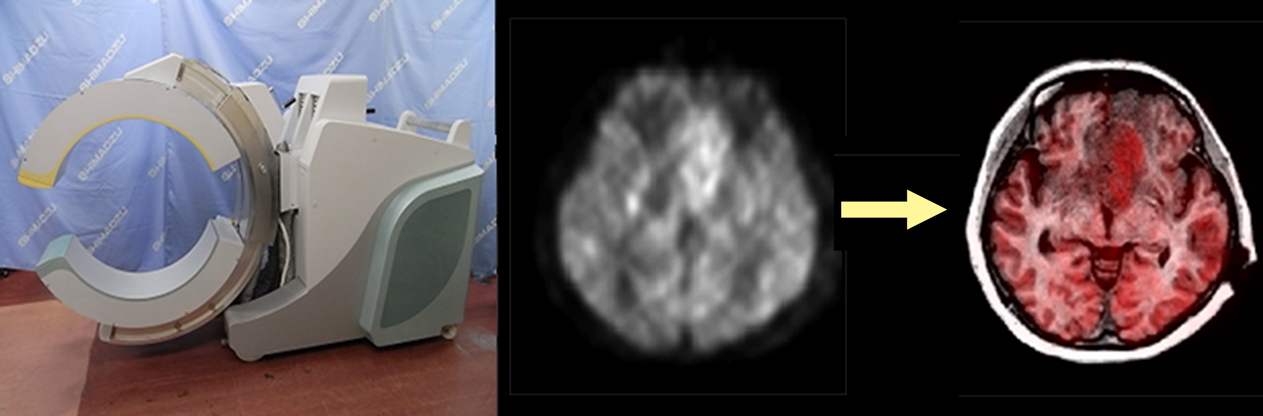
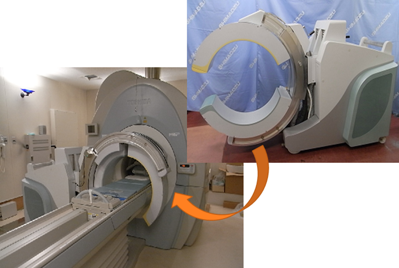
The details of fxPET scanner were already reported, and briefly described as follows: Scanners: Flexible PET (fxPET) is a dual-head mobile PET system, based on a silicon photomultiplier (SiPM)-based depth-of-interaction (DOI) time-of-flight (TOF) detector with MR compatibility (Shimadzu Corporation, Kyoto, Japan) (Fig. 1). FxPET was equipped with an existing MR unit, where 1.5T MR scanner (Excelart Vantage, Toshiba Medical Systems Corporation, Otawara, Japan), thus fxPET/MRI unit realized sequential acquisition of PET and MR images (Fig. 2).
- Protocols
All subjects underwent a single-injection of 18F-fluoro-2-deoxy-D-glucose (FDG). After clinical PET/CT scans, subsequently subjects moved to the fxPET/MRI unit room and underwent fxPET scans followed by MRI scans.
- Image reconstruction
Attenuation maps was created by hybrid segmentation-atlas method (HSAM). In this method, atlas data is used for low signal region of bone and air which is difficult to segment.
Image reconstruction was conducted by two methods: (i) conventional DRAMA algorithm and (ii) DRAMA-NLM.
– DRAMA: F(x) = L(y, Ax) y: observed data, A: system matrix, L:log-likelihood function
– DRAMA-NLM: F(x) = L(y, Ax) + α U(x) α: regularization parameter, U(x): smoothing penalty function using NLM filter.
- Image analysis
Standardized uptake value (SUV) was compared between DRAMA and DRAMA-NLM. PET images were registered to the corresponding 3D T1WI, then the mask of gray matter was applied, and the mean SUV of gray matter was calculated for PET images reconstructed with DRAMA and DRAMA-NLM.
One neuroradiologist subjectively compared PET images reconstructed with DRAMA and DRAMA-NLM.
Results
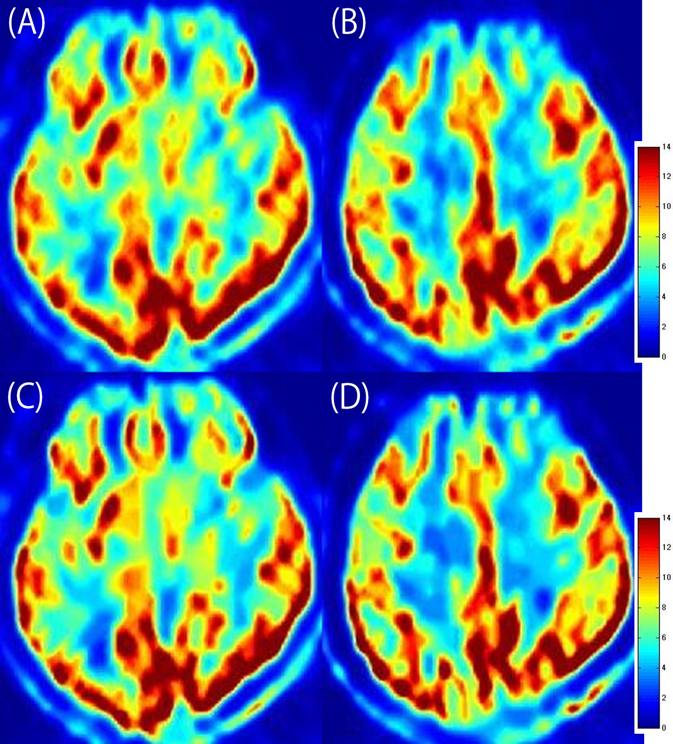
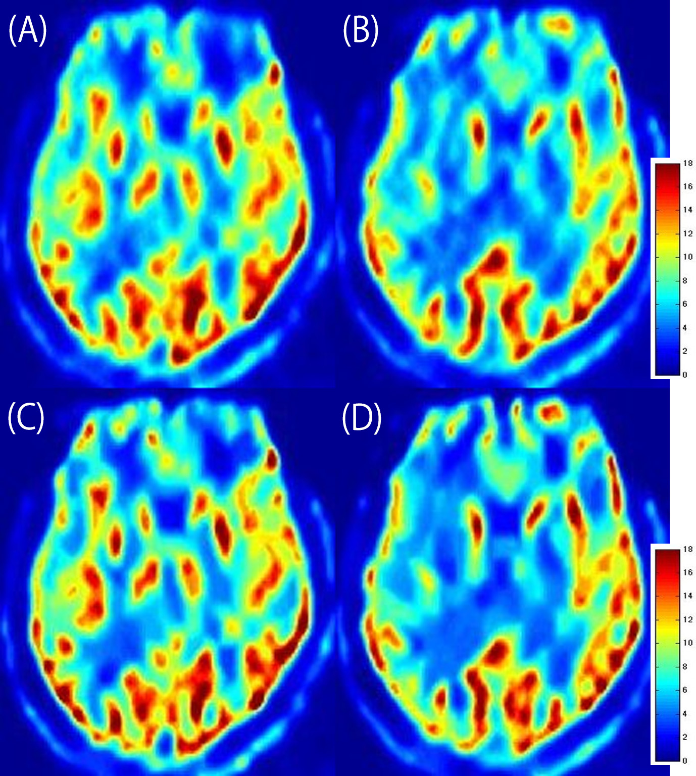
Representative images are shown in Fig .3 and Fig 4.
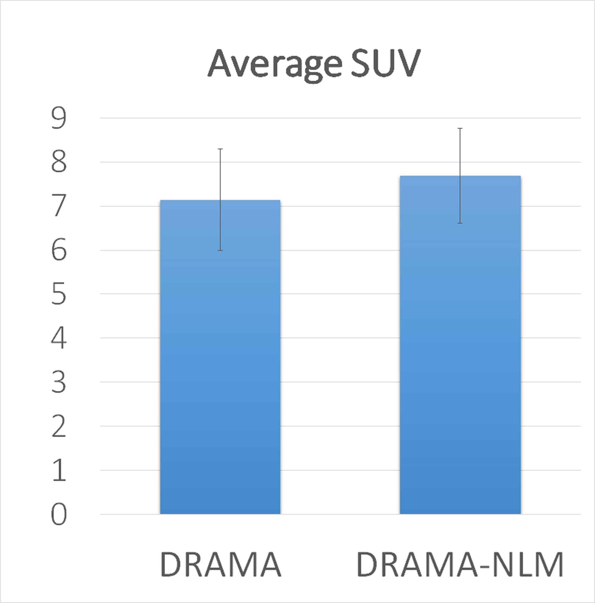
The average SUVs of DRAMA and DRAMA-NLM were 7.14 ± 1.15, 7.67 ± 1.08, respectively(P=0.001) (Fig. 5).
The image quality of fxPET images reconstructed with DRAMA and DRAMA-NLM was almost similar and acceptable, however, DRAMA-NLM showed slightly better demarcated gray matter uptake than DRAMA.
Discussion
Although the image degradation by DRAMA is uncommon in brain fxPET, DRAMA-NLM significantly increase SUV, therefore, DRAMA-NLM contributed to potential improvement of brain fxPET.
Conclusions
This study showed the improvement of brain fxPET image by implementing a newly developed NLM algorithm.Acknowledgements
This study was conducted as a part of the project supported by NEDO (New Energy and Industrial Technology Development Organization).References
1. Tetsuya Kobayashi, Keishi Kitamura Technology Research Laboratory, Shimadzu Corporation, Kyoto, Japan Design considerations for a partial-ring, multi-modal compatible whole-body TOF PET scanner: Flexible PET. Nuclear Science Symposium and Medical Imaging Conference (NSS/MIC), 2012 IEEE DOI: 10.1109/NSSMIC.2012.6551640
2. Masayuki Nakazawa, Junichi Ohi, Tetsuo Furumiya, Tomoaki Tsuda, Masafumi Furuta, Masanobu Sato, Keishi Kitamura, "PET data acquisition (DAQ) system having scalability for the number of detector", Nuclear Science Symposium and Medical Imaging Conference (NSS/MIC) 2012 IEEE, pp. 2475-2478, 2012
3. Masafumi Furuta, Masanobu Satoh, Junichi Ohi, Hiromichi Tonami, Tetsuo Furumiya, Tomoaki Tsuda, Masayuki Nakazawa, Nobuya Hashizume, Tetsuya Kobayashi, Keishi Kitamura, "Development of a proof of concept system for multi-modal compatible PET: Flexible PET", Nuclear Science Symposium and Medical Imaging Conference (NSS/MIC) 2013 IEEE, pp. 1-4, 2013.
4. Yoshiyuki Yamakawa, Tetsuya Kobayashi, Masafumi Furuta, Masanobu Sato, Junichi Ohi, Hiromichi Tonami, Tetsuo Furumiya, Tomoaki Tsuda, Masayuki Nakazawa, Nobuya Hashizume, Keishi Kitamura, "Development of a dual-head mobile DOI-TOF PET system having multi-modality compatibility", Nuclear Science Symposium and Medical Imaging Conference (NSS/MIC) 2014 IEEE, pp. 1-3, 2014.
5. Asuka Tanigawa, Taiga Yamaya, Hiroshi Kawaguchi, Yoshiyuki Hirano, Takahiro Shiraishi, Katsuyuki Tanimoto, Eiji Yoshida, Hiroshi Ito, Takayuki Obata, and Mikio Suga, “Hybrid segmentation-atlas method for PET-MRI attenuation correction”, Nuclear Science Symposium and Medical Imaging Conference (NSS/MIC) 2012 IEEE, pp. 2727-2729, 2012
Figures