1913
Evaluation of Treatment Effect for Saccular Aneurysm by DANTE T1-SPACE1Department of Diagnostic Imaging and Nuclear Medicine, Kyoto University Graduate School of Medicine, Kyoto, Japan, 2Department of Neurosurgery, Kyoto University Graduate School of Medicine, Kyoto, Japan, 3Human Brain Research Center, Kyoto University Graduate School of Medicine, Kyoto, Japan, 4Siemens Healthineers, Portland, OR, United States, 5Siemens Healthineers, San Francisco, CA, United States
Synopsis
The purpose of this study is to evaluate the therapeutic effect by FD stent on DANTE T1-SPACE imaging by comparing contrast enhanced 3D T1-weighted imaging. Patients underwent MR imaging for evaluation of pre-, post FD stent placement, and follow-up at 3T MR scanners were included. DANTE T1-SPACE of aneurysm showed dark intensity in pre-treatment study, and higher intensity in follow-up study, then darker intensity later. Enhancement ratio showed high value in pre-treatment study, and low value in follow-up study. DANTE T1-SPACE of aneurysm and enhancement ratio were negatively associated in all patients and exams.
Introduction
Flow diverter (FD) stent is a new endovascular tool for huge cerebral aneurysms. Highly dense mesh of the FD reduces inflow and outflow on the aneurysm, resulting in intra-aneurysmal thrombosis. The time course of the intra-aneurysmal thrombosis remains unclear, and evaluation strategy is not determined.
DANTE T1-SPACE is a novel image sequence which provides a good black blood imaging by using DANTE pulse train as well as phase dispersion effect of 3D turbo spin echo sequence. T1 SPACE with DANTE preparation, four-fold accelerated by CAIPIRINHA (Controlled Aliasing In Parallel Imaging Results IN Higher Acceleration), can provide high-resolution (0.56 mm isotropic) anatomical imaging within clinically feasible scan time.
The purpose of this study is to evaluate the therapeutic effect by FD stent on DANTE T1-SPACE imaging by comparing contrast enhanced 3D T1-weighted imaging.
Methods
- Subjects
Seven patients who underwent FD stent placement for intracranial aneurysms were enrolled in this study. This study was approved by the institutional review board.
- MR imaging
Patients underwent MR imaging for evaluation of pre-, post FD stent placement, and follow-up at 3T MR scanners (MAGNETOM Skyra or Prisma, Siemens Healthcare GmbH, Erlangen, Germany) with a 32-channel or 64-channel head coil. The imaging protocol included DANTE T1-SPACE and the parameters were as follows: sagittal acquisition; TR/TE, 1000 ms/11 ms; variable flip angle; echo train length, 60; FOV, 180 × 180 mm; resolution, 0.56×0.56×0.56 mm; CAIPIRINHA, acceleration factor of 4; fat suppression; and acquisition time 5 min 44 sec. Pre- and Gd- enhanced T1WI were also performed.
- Post-imaging process and ROI analysis
Post FD stent placement and follow-up DANTE and T1WI were registered to pre-treatment study by SPM12. ROIs were placed in intra-aneurysm (An), Pons for DANTE, pre- and Gd- enhanced T1WI. Signal of DANTE T1-SPACE was evaluated by
An_DANTE/Pons_DANTE.
Enhancement ratio was defined as
An_GdT1/Pons_GdT1 - An_preT1/Pons_preT1.
Results
Representative cases are shown in Fig.1 and Fig. 2.
DANTE T1-SPACE of aneurysm showed dark intensity in pre-treatment study, and higher intensity in follow-up study. About 100 days after FD stent placement, intensity will become darker (Fig. 3).
Enhancement ratio showed high value in pre-treatment study, and low value in follow-up study (Fig. 4).
DANTE T1-SPACE of aneurysm and enhancement ratio were negatively associated in all patients and exams (R² = 0.72).
Discussion
DANTE T1-SPACE visualize the status of thrombus formation inside the aneurysm of aneurysm thanks to its high-resolution information as black blood imaging. Thrombus shows high intensity post-stent placement study, subsequently decrease in signal intensity at chronic state in follow-up study.
Contrast enhanced T1WI is necessary for the precise evaluation of the relationship between FD stent and thrombus formation. Because high correlation is seen between signal of DANTE T1-SPACE and enhancement ratio, DANTE T1-SPACE may substitute contrast enhanced MRI, especially in follow-up study.
Relatively small number of patients were included in this study, which is one of limitations. DANTE T1-SPACE of aneurysm and enhancement ratio may be evaluated in a larger number of patients.
Acknowledgements
We are grateful to Mr. Katsutoshi Murata, Mr. Yuta Urushibata, and Mr. Hirokazu Kawaguchi, Siemens Healthcare Japan K. K. for their kind help.References
1. Li L, Miller KL, Jezzard P. DANTE-prepared pulse trains: a novel approach to motion-sensitized and motion-suppressed quantitative magnetic resonance imaging. Magn Reson Med. 2012 Nov;68(5):1423-38.
2. Li L, Chai JT, Biasiolli L, Robson MD, Choudhury RP, Handa AI, Near J, Jezzard P. Black-blood multicontrast imaging of carotid arteries with DANTE-prepared 2D and 3D MR imaging. Radiology. 2014 Nov;273(2):560-9
3. Xie Y, Yang Q, Xie G, Pang J, Fan Z, Li D. Improved black-blood imaging using DANTE-SPACE for simultaneous carotid and intracranial vessel wall evaluation. Magn Reson Med. 2016 Jun;75(6):2286-94
Figures
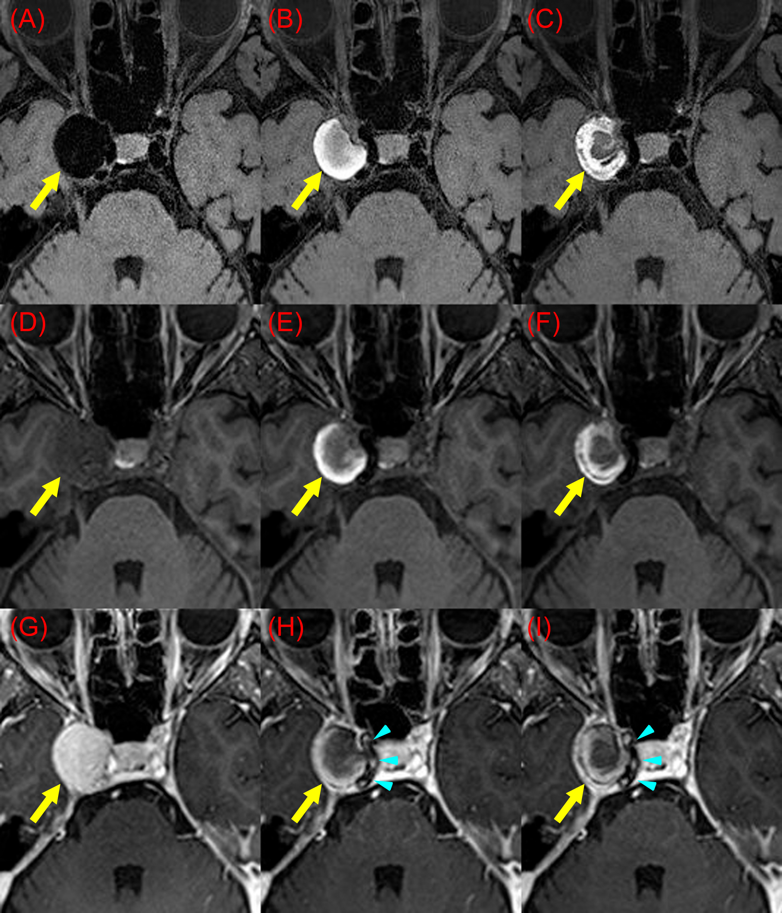
Fig. 1.
A huge right interanal carotid aneurysm (yellow arrows, Patient 5). DANTE T1-SPACE, (A), (B), and (C); Pre-contrast T1WI, (D), (E), and (F); Gd-enhanced T1WI, (G), (H), and (I). Pre-treatment study shows very low signal on DANTE T1-SPACE (A), slightly low signal on Pre-contrast T1WI (D), and good enhancement (G). Post FD stent placement study (day 35, B, E, H) and follow-up study (day 73, C, F, I) shows high signal on DANTE T1-SPACE and pre-contrast T1WI, and no apparent enhancement on Gd-T1WI. Blue arrowheads indicate FD stent. Blue arrowheads indicate FD stent.
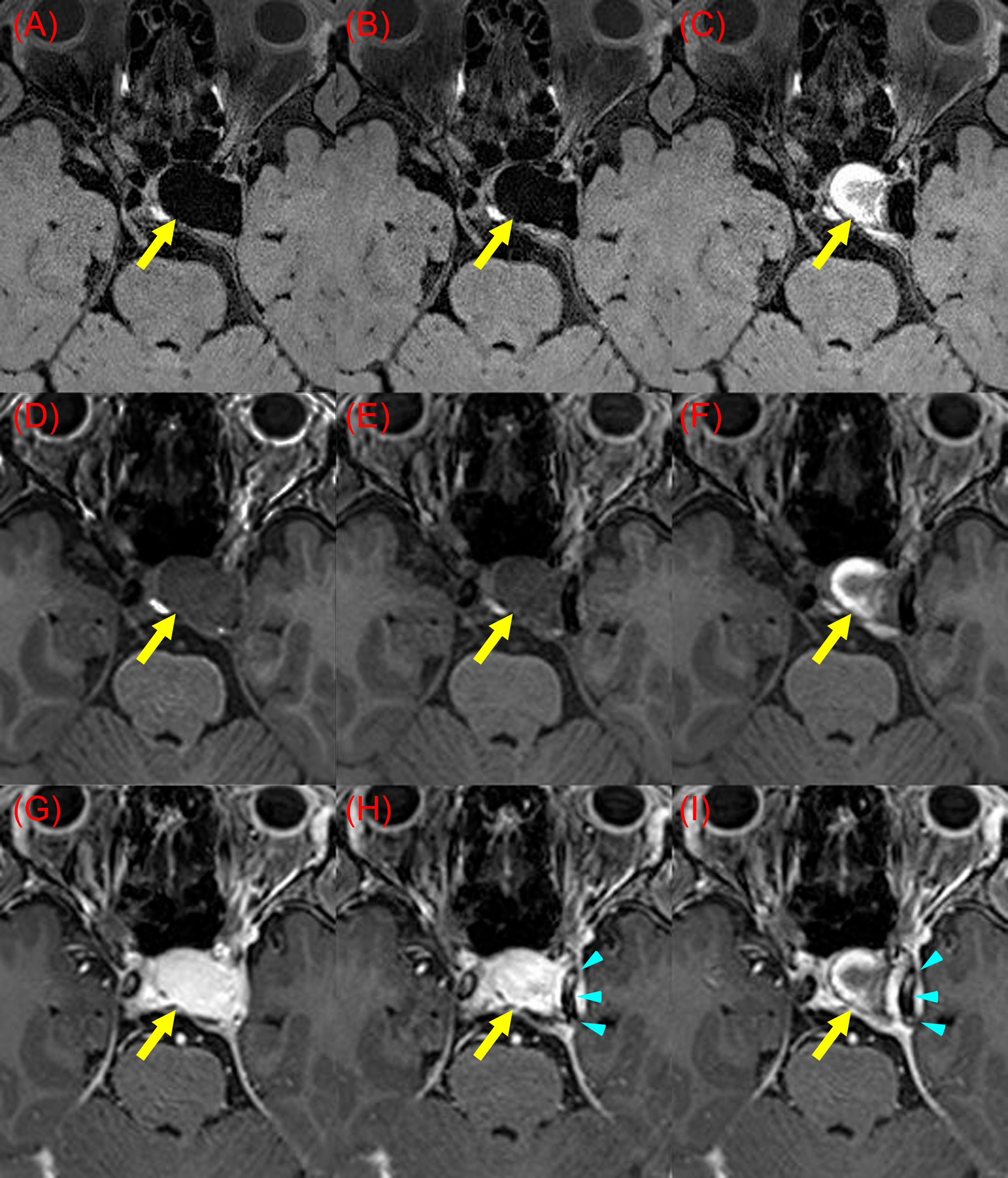
Fig. 2.
A huge left interanal carotid aneurysm (yellow arrows, Patient 4). DANTE T1-SPACE, (A), (B), and (C); Pre-contrast T1WI, (D), (E), and (F); Gd-enhanced T1WI, (G), (H), and (I). Pre-treatment study shows very low signal on DANTE T1-SPACE (A), slightly low signal on Pre-contrast T1WI (D), and good enhancement (G). Post FD stent placement study shows similar findings as pre-treatments study except slight thrombus formation (B, E, H). Follow-up study shows high signal on DANTE T1-SPACE (C) and pre-contrast T1WI (F), and no apparent enhancement on Gd-T1WI (I). Blue arrowheads indicate FD stent.
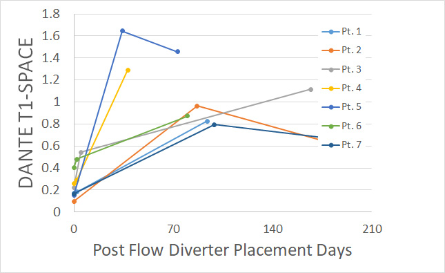
Fig. 3.
DANTE T1-SPACE of aneurysm showed dark intensity in pre-treatment study, and higher intensity in follow-up study. About 100 days after FD stent placement, intensity will become darker.
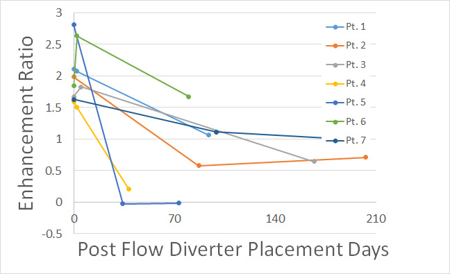
Fig. 4.
Enhancement ratio showed high value in pre-treatment study, and low value in follow-up study.
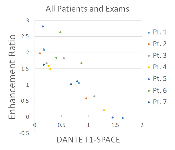
Fig. 5.
Signal of DANTE T1-SPACE inside the aneurysm and enhancement ratio were negatively associated in all patients and exams (R² = 0.72).