1906
The value of diffusion tensor imaging (DTI) and tractography (DTT) in lumbar nerve roots display and lumbar disc herniation assessment1The first affiliated hospital of Dalian medical university, Dalian, China, 2GE Healthcare, Beijing, China
Synopsis
This is a prospective study on lumbar disc herniation patient and healthy control with diffusion tensor imaging (DTI) and tractography (DTT). We obtained a high success rate (>90%) of achieving the DTI with tractography of lumbar nerve roots was in this study, and revealed that DTI and DTT technique can both display intensity and morphology changes in the compressed areas of lumbar nerve roots. DTI with tractography provides an abundant diagnostic information with specificity on both qualitative- and quantitative-wise, which is great helpful to assess the disorders with lumbar nerve root compression.
Purpose
To evaluate the DTI and DTT of lumbar nerve roots in the diagnosis of lumbar disc herniation through all the related qualitative info from tractography and quantitative measures of the fraction anisotropy (FA) value and apparent diffusion coefficient (ADC) value.
Methods and materials
This prospective study was approved by our Institutional Review Board and written informed consent was obtained from all attendees. Twenty patients (age=27-67 years; 10 males, 10 females) with clinically confirmed lumbar disc herniation (left: 11; right: 9) and 20 normal controls (age=26-63 years; 10 males, 10 females) were performed DTI, DTT and axial T2W MRI scanning on a GE Signa HDxt 3.0T MR scanner. Previous history of spinal trauma, surgery or neurological diseases was exclusion criteria for both groups. Fiber tracking images were set from lumbar nerve roots in axial DTI images and fused with T2WI image as an anatomic background. The FA value and ADC value of left- and right-side nerve roots were measured in both groups. The difference between groups was compared with t-test.Results
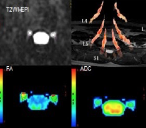
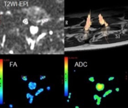
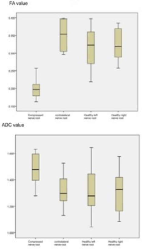
A high success rate (>90%) of achieving the DTI with tractography of lumbar nerve roots was obtained in this study (Fig. 1). At the compressed areas of the lumbar nerve roots, a high signal intenstity was observed in the T2 images of DTI from the patients comparing with the contralateral and from the controls. In addition, apparent morphological changes were also observed at the corresponding regions in a pattern of shift, bending, sparsity in number and so on (Fig. 2). No significant difference in the mean FA and ADC values was observed between the left- and right-side nerve roots at same level (L5 and S1) and in between. However, the mean FA values of compressed nerve roots were statistically lower (p<0.05) than the contralateral and the controls, 0.197±0.032 vs. 0.351±0.044; and the mean ADC values of compressed nerve roots were significantly higher (p<0.05) than the contralateral and the controls, (1.48±0.13) ×10-3 mm2/s vs. (1.32±0.14) ×10-3 mm2/s.(Fig.3)Discussion and Conclusion
Eguchi [1] used diffusion tensor and fiber tractography technology successfully tracked human lumbar spinal nerve, not only applicable to normal, as well as for patients with lumbar disc adaptation. Our study also confirms this, and the tractography images were fused with T2WI sequences as anatomy background for a better display of process and morphology of nerve roots. DTI with tractography provides an abundant diagnostic information with specificity on both qualitative- and quantitative-wise, which is great helpful to assess the disorders with lumbar nerve root compression, e.g. lumbar disc herniation.Acknowledgements
References
[1]. Y. Eguchi S, Ohtori S, Orita H, et al.Quantitative evaluation and visualization of lumbar foraminal nerve root entrapment by using diffusion tensor imaging: preliminary results. Am J Neuroradiol. 2011; 32(10):1824-1829.Figures