1905
Language reorganization in pre-and post-operative drug refractory extra temporal lobe epilepsy patients: An fMRI based study1Department of Neurology, All India Institute of Medical Sciences, New Delhi, India, 2Department of NMR and MRI Facility, All India Institute of Medical Sciences, New Delhi, India, 3Department of Neuro-Surgery, All India Institute of Medical Sciences, New Delhi, India, 4Department of Clinical Neuropsychology, All India Institute of Medical Sciences, New Delhi, India
Synopsis
Drug refractory epilepsy (DRE) patients have atypical language lateralization with ipsilateral and contra lateral hemispheric lesions and pathological abnormalities. Such kind of patients may have different language recovery after surgery. In this study, we have used a standardized Hindi-language paradigm using semantic, syntactic, judgement and comprehension components for testing in the North-Indian population. We observed greater improvement in language skills in ETLE-patients with correspondingly greater recruitment of the bilateral hemisphere.
Introduction
Drug refractory epilepsy has a prevalence of ~30% of people with epilepsy in India, [1-2] and may have associated cognitive deficits, including impairment of language (78%) and executive functions (55%).[2] In patients with epilepsy, language lateralization is more often atypical due to the influence of interictal discharges and seizure spread. [2] In extra temporal lobe epilepsy (ETLE) patients with frontal lobe lesion, some of the language components are observed to be affected [5]. Our aim was to determine whether there were differences in the extent of improvement and the pattern of reorganization of language functions on functional magnetic resonance imaging (fMRI) in refractory ETLE after epilepsy surgeryMethod
Consecutive 20 ETLE patients (mean age 26.85±5.4 years, 13M / 7F) with left hemispheric language dominance (12 with left frontal lesion; 4 with left parieto-temporal lesion; 2 subjects with left parieto-occipital lesion and 2 patients with left posterior occipito-temporal lesion) and 24 healthy controls (mean age 27± 6.7 years, 18M / 6F) were recruited for the study. Ten (out of 20) ETLE patients underwent two sessions of clinical assessments and fMRI, i.e., both before and after an average of 6.61 (± 1.85) months of surgery. Multiple aspects of language (repetition, naming, word fluency, visual word and comprehension reading) were tested using the Speech Language dysfunction (SLD) and Visual reading dysfunction (VRD) modules of Indian Aphasia Battery (IAB) in the Hindi language. FMRI was performed using a standardized Hindi language paradigm (lexical, semantic, syntactic and comprehension components) in both cases and controls, before and after epilepsy surgery (in cases) at 1.5 T MR Scanner using visual cues projecting through MR compatible LCD goggles (NNL, Norway) mounted on 8 channel head coil. Single-shot echo planar imaging (EPI) sequence was used for the BOLD studies with parameters: number of slices 29, slice thickness 4.5 mm, slice gap 0 mm, echo train length 63, repetition time 2000 ms, echo time 24 ms, field of view: 230 mm, resolution: 64x64 and total number of measurements 251. Data analysis and group comparisons were carried out using SPM8.Results and discussion
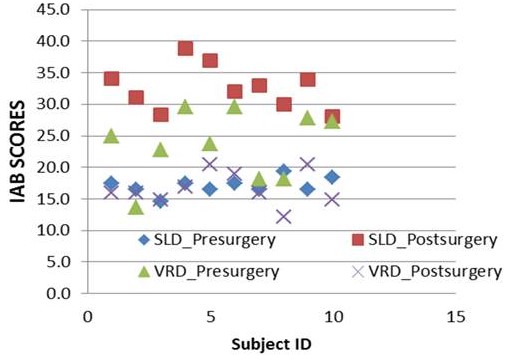
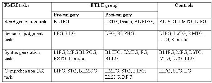
At baseline, clinical testing with IAB exhibited better scores in controls than in patients. Clinical SLD and VRD scores revealed significant improvement in the ETLE postoperatively (Figure 1). All patients were seizure free after surgery. Blood-oxygen-level dependent (BOLD) activation was observed in bilateral cerebral regions (Table 1) during language tasks in the ETLE cases, while it was in the left hemispheric traditional language areas in controls. Atypical BOLD activation in pre-operative session of ETLE patients may be attributed to long duration of epilepsy and frequent seizures originating from the left hemisphere.
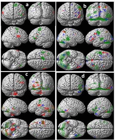
Post-operatively, greater BOLD activation was observed in the bilateral inferior frontal gyri (IFG, r =0.45*; p<0.05), middle frontal gyrus (MFG, r =0.67**; p<0.01) superior temporal gyrus (STG, r =0.78* p<0.02) and angular gyrus (AG, r =0.73*; p<0.04) in comparison to that of pre-surgery (Figure 2, Table 1). BOLD activations in IFG, STG and AG areas were attributed to lexical, semantic and syntactic information proceeding during language task [3-4] and were not prominent in pre-surgery patients group. Recruitment of right hemispheric BOLD activation suggests language reorganization due to impaired language networks [4]. Our results suggest post-operative language restoration in ETLE patients, similar to earlier studies exhibiting improvement in language function in patients with DRE after surgery with functional reorganization [3-4]. Since the location of the lesion varies in ETLE patients, further studies may be required to clarify the role of the language function in these patients.
Conclusion
Greater improvement in language skills in ETLE-patients with correspondingly greater recruitment of the bilateral hemisphere, becoming similar to that of healthy controls after epilepsy surgery suggest functional reorganization of language areas.Acknowledgements
No acknowledgement found.References
1. Sherman E et al., 2011 Epilepsia, 52, 857–869.
2. Wang W. et al., 2011 Epilepsy Behav. 22, 728-734.
3. Wong S et al., Neurology 2009; 73: 518-525.
4. Chaudhary K et al., 2014 Indian Journal of Radiology & Imaging; 24: 51-56.
5. Zhang et al., 2013 Neuropsychiatric Disease and Treatment; 9, 1673–1682.
Figures