1903
Quantitative Susceptibility Mapping analysis of cerebral microbleeds in hypertensive patients1The 4th center hospital of TianJin, China, TianJin, China, 2Philips Healthcare Beijing China, Beijing, China
Synopsis
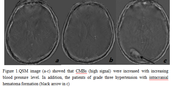
Cerebral microbleeds (CMBs) was often found in hypertensive patients.Quantitative susceptibility mapping (QSM) could detect iron-containing lesions with high sensitivity and spatial accuracy in the presence of potentially confounding tissue abnormalities.The results of retrospective study showed that there was significant difference in CMBs between the hypertensive group and the control group.So the conclussion is MR quantitative susceptibility could directly explicate the evolution law of CMBs in hypertensive patients, timely intervention of hypertension could reduce the occurrence of CMBs.
INTRODUCTION
Cerebral microbleeds (CMBs) was often found in hypertensive patients.Histopathology confirmed that microbleeds was due to micro-arterial fiber hyaline degeneration and micro-extravasation of blood caused by hemosiderin deposition1,2. Quantitative susceptibility mapping (QSM) could detect iron-containing lesions with high sensitivity and spatial accuracy in the presence of potentially confounding tissue abnormalities3. A retrospective study was performed to explore the risk factors of cerebral microbleed in hypertensive patients and the relevance of cerebral microbleeds with hypertension.METHODS
15 healthy volunteers and 15 hypertensive patients were include in this study. According to WHO guidelines for the treatment of hypertension, hypertensive patients are divided into 7 cases of grade one hypertension, 5 cases of grade two hypertension, 3 cases of grade three hypertension.And the study was approved by the ethics committee of Tianjin Forth centre Hospital, Tianjin, China. High resolution 3D TFE high images were obtained with a 3.0T MR scanner (Achieva TX, Philips Healthcare, Best, the Netherlands). The parameters of the single-echo 3D TFE acquisition were: TE=3.8ms, TR=8.3ms, flip angle=10, FOV =220 x 200 x 120mm, echoes=1 and slice thickness =1.0mm. The QSM processing was performed by using Stisuite tollbox (Version 3.0), to obtain the susceptibility values for all regions of intereston (ROI) based on the amplitude and phase images,the range of ROI is consistent with the high signal in the phase images.All statistical evaluations were performed with software (SPSS 17.0, Armonk, New York, USA) and p<0.05 was considered to indicate a significant difference. Pearson’s correlation was performed to investigate the relationship between the lesion magnetic susceptibility and systolic pressure.RESULTS
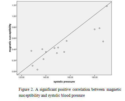
The MR quantitative susceptibility of CMBs in hypertensive group were (1.50±0.32) ppm while control group were (0.75±0.19) ppm, which had a significant difference ( t=3.59,P<0.01). There was a significant difference in susceptibility between the different grades of hypertension (F=19.62, P <0.01). The magnetic susceptibility is positively correlated with the systolic pressure (r=0.85, P <0.01).DISCUSSION
The results showed that the incidence of CMBs in hypertensive patients was significantly higher than the control group, hypertension was an important factor in small arterial lipid-like changes and micro-aneurysms leading to CMBs4. CMBs could be understood as a type of target organ damage in the brain of chronic hypertension5. The results were consistent with the previous studies6 which shown that the severity of CMBs were positively correlated with systolic pressure. The reason is that the severity of microvascular injury is closely related to blood pressure levels and duration.CONCLUSION
MR quantitative susceptibility could directly explicate the evolution law of CMBs in hypertensive patients, timely intervention of hypertension could reduce the occurrence of CMBs.Acknowledgements
I′d like to express my appreciation to the professors and staff of the fourth central hospital for their participation in the study,thanks a lot for your strong support from Phlips.References
1. Greenberg SM, Vernooij MW, Cordonnier C, et al. Cerebral microbleeds: a guide to detection and interpretation[J]. Lancet Neurol,2009 ;8 (2): 165 – 174.
2. Vernooij MW, vander Lugt A, Ikram MA, et al. Prevalence and risk factors of cerebral microbleeds: the Rotterdam Scan Study [J].Neurology,2008; 70 (14): 1208 –1214.
3. Tian Liu, Krishna Surapaneni , MPMin Lou,et al.Cerebral Microbleeds:Burden Assessment by Using Quantitative Susceptibility Mapping[J].Radiology,2012; 262:269 – 278.
4. Wardlaw JM, Lewis SC,Keir SL,et al. Cerbral microbleeds areassociated with lacunar stroke defined clinically and radiologically,independently of white matter lesions[J].Stroke,2006;37(10):2633-2636.
5. Lee SH,Park JM,Kwon,SJ,et al.Left ventricular hypertrophy is associated with cerebral microbleeds in hypentensive pations[J].Neurology,2004;63(1):16-21.
6. Mciiduff CE, Rutkove SB. Critical appraisal of the use of alpha lipoic acid (thioctic acid) in the treatment of symptomantic diabetid polyneuropathy[J].Ther clin risk manag,2011;7:377-385.
Figures