1900
Remote effects of hemodynamic impairment on network efficiency in chronic steno-occlusive disease of the anterior circulation: A resting-state functional MRI studyJunjie Wu1, Seena Dehkharghani2, Fadi Nahab3, Jason W. Allen1, Ranliang Hu1, and Deqiang Qiu1
1Department of Radiology and Imaging Sciences, Emory University School of Medicine, Atlanta, GA, United States, 2Department of Radiology, New York University, New York, NY, United States, 3Department of Neurology, Emory University School of Medicine, Atlanta, GA, United States
Synopsis
In this abstract we explored remote effects of cerebrovascular hemodynamic impairment on the efficiency of functional connectivity in patients with chronic, anterior circulation steno-occlusive disease. We further evaluated the correlation between network efficiency and cerebrovascular reactivity (CVR), a measure of cerebral hemodynamics.
Introduction
Patients with chronic steno-occlusive disease of the cerebrovascular system are at risk of ischemic stroke as well as cognitive decline. Functional connectivity mapping has advanced the understanding of neural mechanisms underlying cognitive impairment. While functional network reorganization has been found in stroke and cerebrovascular disease1-3, little is known about remote effects of hemodynamic compromise on network topology of distant brain areas, such as the hemisphere contralateral to vessel occlusion/stenosis. To address this gap, we aimed to evaluate the efficiency of functional connectivity in the contralateral hemisphere among patients with unilateral chronic steno-occlusive disease, and to investigate the relationship between network efficiency and CVR, a measure of cerebral hemodynamic reserve.Methods
Twenty-one patients with unilateral chronic steno-occlusive disease of the anterior circulation (age, 48.43 ± 14.46 years; 16 females) and twenty age-matched healthy controls (age, 40.75 ± 16.21 years; 8 females) were included. All patients underwent a 20-minute blood oxygen level–dependent (BOLD) acquisition using a gradient-echo echo-planar imaging (EPI) sequence (TR/TE = 2000/30 ms, FA = 78°, voxel size = 3.44 × 3.44 × 4 mm3, 30 slices). After 5 minutes of baseline BOLD acquisition, acetazolamide (ACZ) was slowly infused over 3-5 minutes. CVR was calculated as percentage augmentation of BOLD signals between pre- and post-ACZ infusion. Similarly, BOLD images were acquired from the healthy controls for 13.33 minutes, but without the administration of ACZ. The first 5 minutes of data from both patients and healthy participants were used for network analysis. To calculate graph-theory metrics, forty-five regions of interest (ROIs) were defined according to the Automated Anatomical Labeling atlas4 for each hemisphere. Network efficiency was quantified in terms of the local efficiency Elocal and the nodal efficiency Enodal. In the healthy controls, the mean and standard deviation of network efficiency were calculated for both left and right hemispheres separately as a reference standard for the patients. To quantify abnormality in the hemisphere contralateral to cerebrovascular disease in each patient, network efficiency was scored in terms of z-value relative to the mean and standard deviation of the respective hemisphere of the healthy controls calculated above. The correlation between network efficiency and ROI-specific CVR was evaluated using a mixed-effects model. Subject-specific slope and intercept were modeled as random effects to assess the linear relationship.Results
In general, both Elocal and Enodal were lower in the patients as compared with the healthy controls (Fig 1A and B). Fig 2 shows the z-maps of Elocal and Enodal of the patients relative to the controls. Negative z-scores were observed in most of the ROIs, suggesting compromised network efficiency in the patient cohort. One-sample t-test showed that z-scores of network efficiency were significantly lower than zero at different sparsity levels (20%, 30% and 40%) (Table 1). Visual inspection suggested an association between network efficiency and CVR (Fig 1). Mixed-effects model showed significant correlation between Enodal and CVR (P = 0.009), while no significant correlation was observed between Elocal and CVR (P = 0.976) (Fig 3).Discussion and Conclusion
Our study provides evidence for altered functional connectivity distant from injured sites5. We found reduced network efficiency in the contralateral hemisphere among patients with chronic cerebrovascular disease. Although we focused on the apparently normal hemisphere in this study, its hemodynamic response function might still be altered, contributing to the compromised topological properties. Further studies using simultaneous EEG-fMRI may shed further light on the physiological basis of brain functional networks.Acknowledgements
No acknowledgement found.References
- Chang T-Y, Huang K-L, Ho M-Y, Ho P-S, Chang C-H, Liu C-H, et al. Graph theoretical analysis of functional networks and its relationship to cognitive decline in patients with carotid stenosis. Journal of Cerebral Blood Flow & Metabolism. 2016;36:808-818.
- Wang L, Yu C, Chen H, Qin W, He Y, Fan F, et al. Dynamic functional reorganization of the motor execution network after stroke. Brain. 2010;133:1224-1238.
- Gratton C, Nomura EM, Pérez F, D'Esposito M. Focal brain lesions to critical locations cause widespread disruption of the modular organization of the brain. Journal of Cognitive Neuroscience. 2012;24:1275-1285.
- Tzourio-Mazoyer N, Landeau B, Papathanassiou D, Crivello F, Etard O, Delcroix N, et al. Automated anatomical labeling of activations in spm using a macroscopic anatomical parcellation of the mni mri single-subject brain. NeuroImage. 2002;15:273-289.
- Carrera E, Tononi G. Diaschisis: Past, present, future. Brain. 2014;137:2408-2422.
Figures
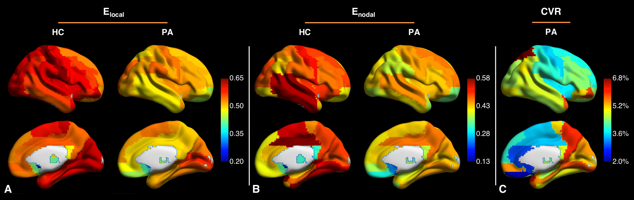
Fig 1. Averaged maps of the local efficiency Elocal (A) and the nodal efficiency Enodal (B) in the right hemisphere of the healthy controls (HC) and in the contralateral hemisphere of the patients (PA). Cerebrovascular reactivity (CVR) map in the contralateral hemisphere of the patients (C). The maps of the patients with right-side disease were left-right flipped to create cohort-averaged maps. Compared to the healthy controls, both Elocal and Enodal were lower among patients.
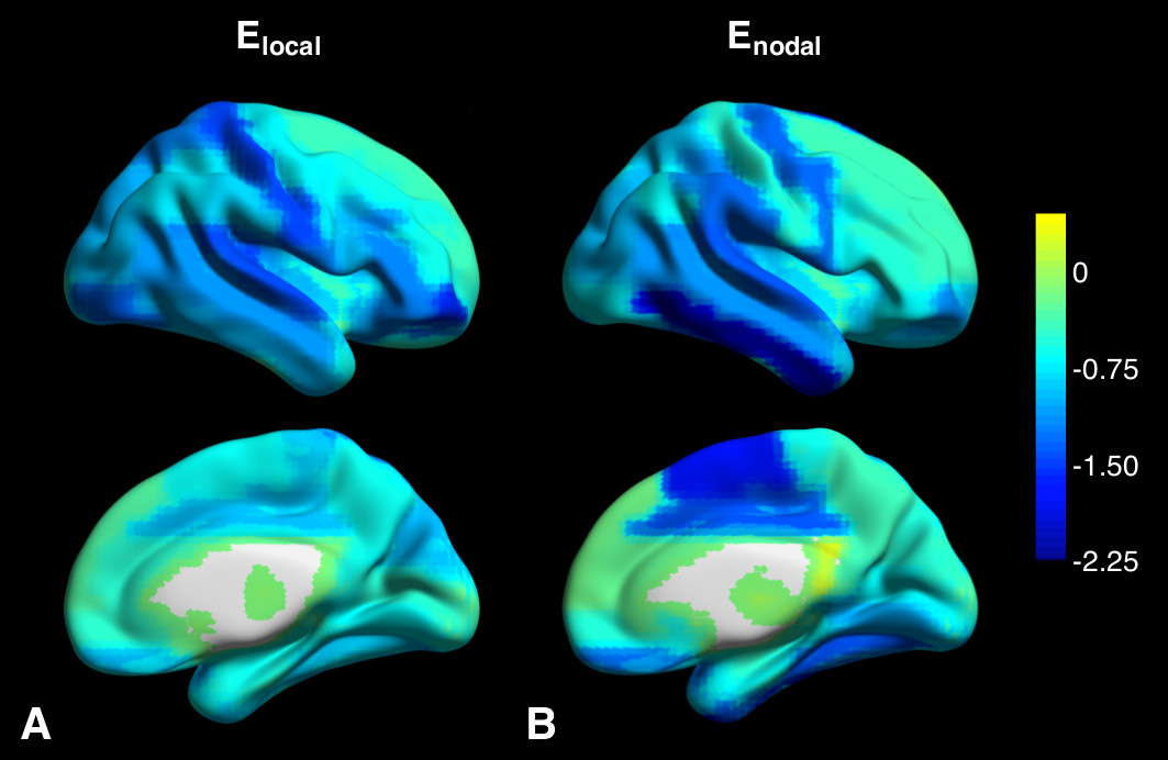
Fig
2. Z-maps of the
local efficiency Elocal
(A) and the nodal efficiency Enodal
(B) in the contralateral hemisphere averaged across all patients. The maps of
the patients with right-side disease were left-right flipped to create cohort-averaged
maps. In most of the brain regions, negative z-scores were found, indicating
lower network efficiency in the patients compared to the healthy controls.
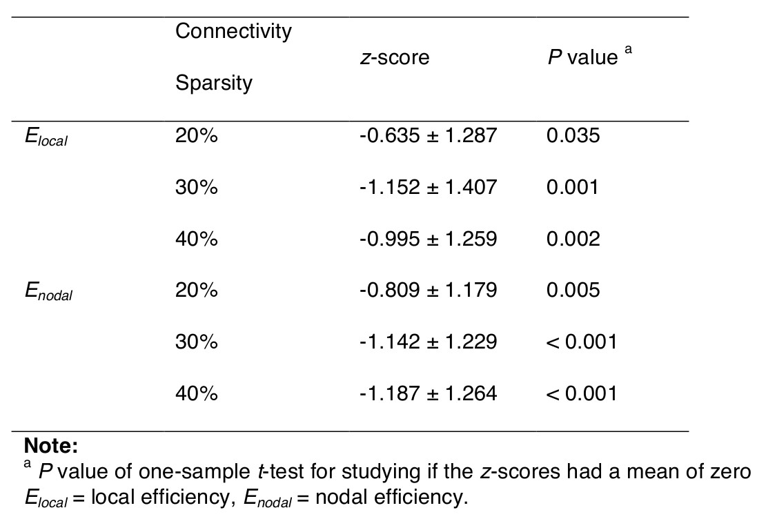
Table
1. Mean z-scores of network efficiency in the
contralateral hemisphere. Z-scores of network efficiency were significantly
lower than zero at different sparsity levels.
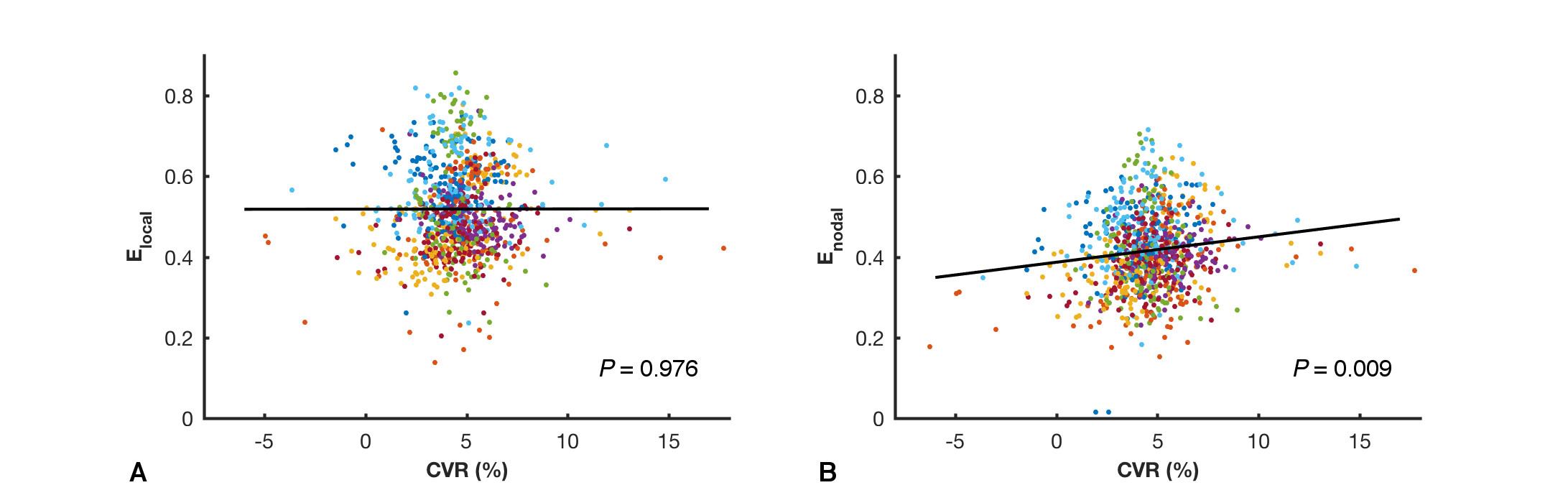
Fig
3. Correlations
between the local efficiency Elocal
and cerebrovascular reactivity (CVR) (A), as well as between the nodal
efficiency Enodal and CVR
(B). Mixed-effects model showed statistically significant correlation between Enodal and CVR (P = 0.009), while no significance was
found between Elocal and
CVR (P = 0.976). Dots of each color
represent a patient. The fitting lines are also shown to indicate trends.