1892
Intravascular Signal Suppression and Micro-Vascular Signal Mapping obtained from ASL Perfusion Imaging with DANTE Pulse1Department of Medical Imaging, Faculty of Life Sciences, Kumamoto University, Kumamoto, Japan, 2Radiology, University of Fukui, Fukui, Japan, 3Radiological Center, University of Fukui Hospital, Fukui, Japan, 4Global MR Applications and Workflow, GE Healthcare Japan, Tokyo, Japan, 5Division of Ultrahigh Field MRI, Institute for Biomedical Science, Iwate Medical University, Iwate, Japan, 6GE Healthcare, Calgary, Canada
Synopsis
In ASL perfusion imaging, the signal from the
label that is still present in larger arteries at the time of imaging causes
vascular artifact, which reduces the accuracy of quantification of cerebral
blood flow. The purpose of this study is to eliminate the vascular artifacts in
larger vessels using the delays alternating with nutation for tailored
excitation (DANTE) pulse as vascular crushing gradients and to evaluate the
efficiency of the DANTE pulse. The optimized DANTE pulse makes it possible to
suppress the vascular signal depending on the flow velocity, which decreased the ASL
signal of the arterial region. The
relative vascular signal mapping may be helpful to reveal altered hemodynamic
state, since the amount of suppressed signal directly associate with flow
velocity.
PURPOSE
Arterial spin labeling (ASL) is a method of obtaining noninvasive measurements for brain perfusion. The signal from the label that is still present in larger arteries at the time of imaging causes vascular artifact as a high signal intensity. This artifact reduces the accuracy of quantification of the arterial transit time and cerebral blood flow. Therefore, it is necessary to clarify which ASL signal originates from a large vessel or microvasculature. The purpose of this study is to eliminate the vascular artifacts in larger vessels using the delays alternating with nutation for tailored excitation (DANTE) pulse as vascular crushing gradients and to evaluate the efficiency of the DANTE pulse.METHODS
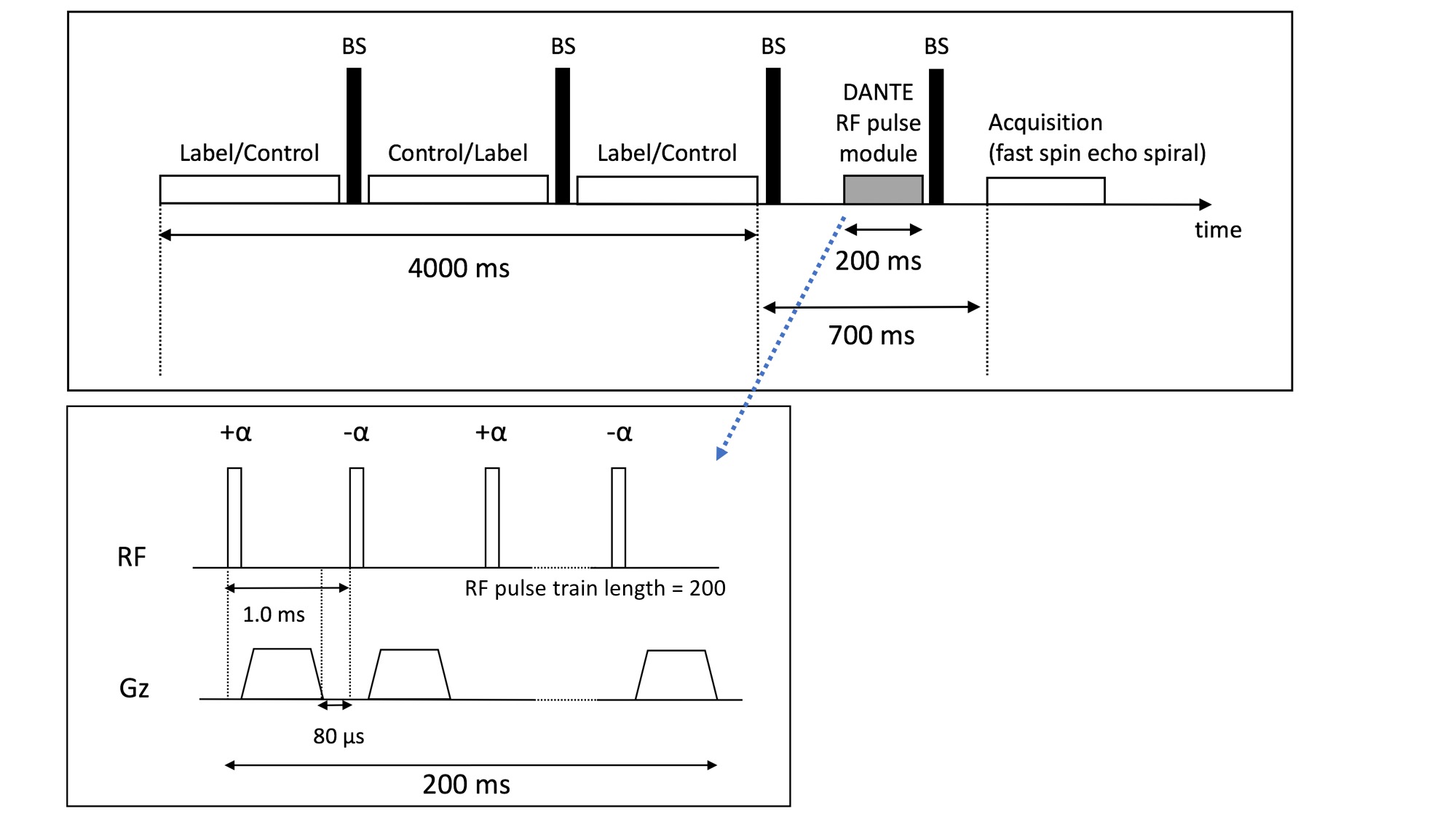
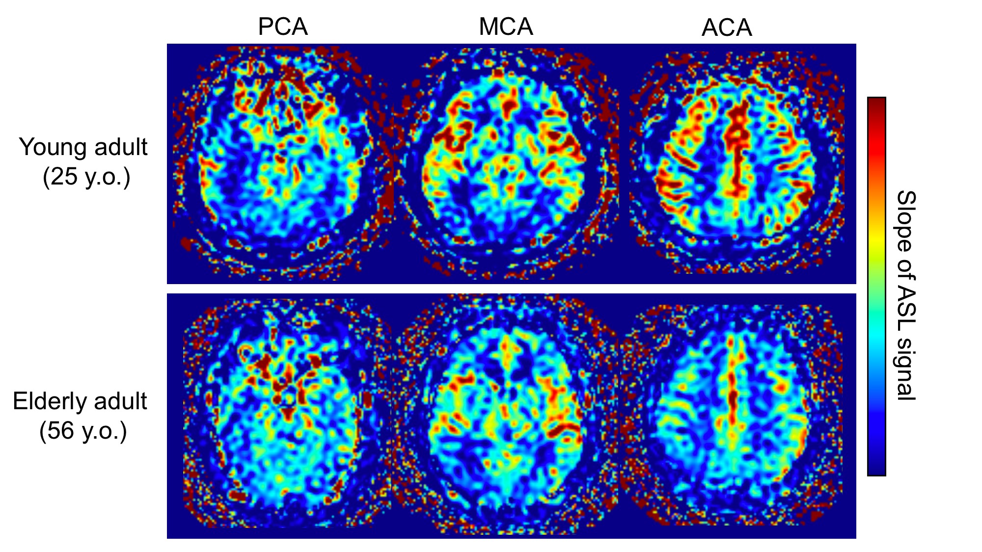
All the studies were performed on a 3.0 T MRI (Discovery MR750; GE Healthcare) with a 32-channel coil. The study protocol was approved by the institutional review board of the University of Fukui. The perfusion phantom, which consists of a labeling and perfusion section, was used. Ten healthy volunteers were enrolled, from which five were categorized in a group of young adults and the other five in a group of elderly adults. All perfusion images acquired pseudo-continuous labeling with three-dimensional FSE with spiral acquisition. The DANTE and ASL sequence are shown in Fig. 1. The RF pulse train of the DANTE was a rapid series of low flip angle (FA) with nonselective RF pulses and gradient pulses. The gradient of the DANTE was applied in the static magnetic field direction along with the direction of the blood vessel in the brain. The gradient of the DANTE was adjusted by the gradient area (GA) defined by multiplying gradient strength and duration. To estimate the optimal FA of the DANTE, static and flowing magnetization were simulated after the DANTE. Then, to evaluate the impact of the FA and the GA of the DANTE, ASL images were obtained with three flow velocity and various parameters of the DANTE using the phantom. The flow settings and parameter of the DANTE were as follows: flow velocity in the imaging area: 2.0, 4.2, 5.7 cm/s; FA: 0-15 degrees; GA: 0-20 μs・T/m, respectively. The imaging parameters of the ASL were as follows: labeling duration, 4.0 s; post-labeling delay time, 700 ms. Finally, to validate the optimal DANTE parameters, volunteers' images were obtained with and without the DANTE under the same conditions with the phantom study. We measured ΔM in each arterial territory: anterior cerebral artery, middle cerebral artery, and posterior cerebral artery in all the volunteers’ images. Moreover, the ratio of ΔM decrease associated with an increased FA in the condition of GA = 10 μs・T/m was measured and its slope was calculated. This reduction of ASL signal should be directly associates with the velocity and the amount of microvascular signal. Therefore the mapping of this slope of ASL signal may reveal the relative microvascular space or labeled spin velocity. We compared these images between young and elderly group subjects.
RESULTS
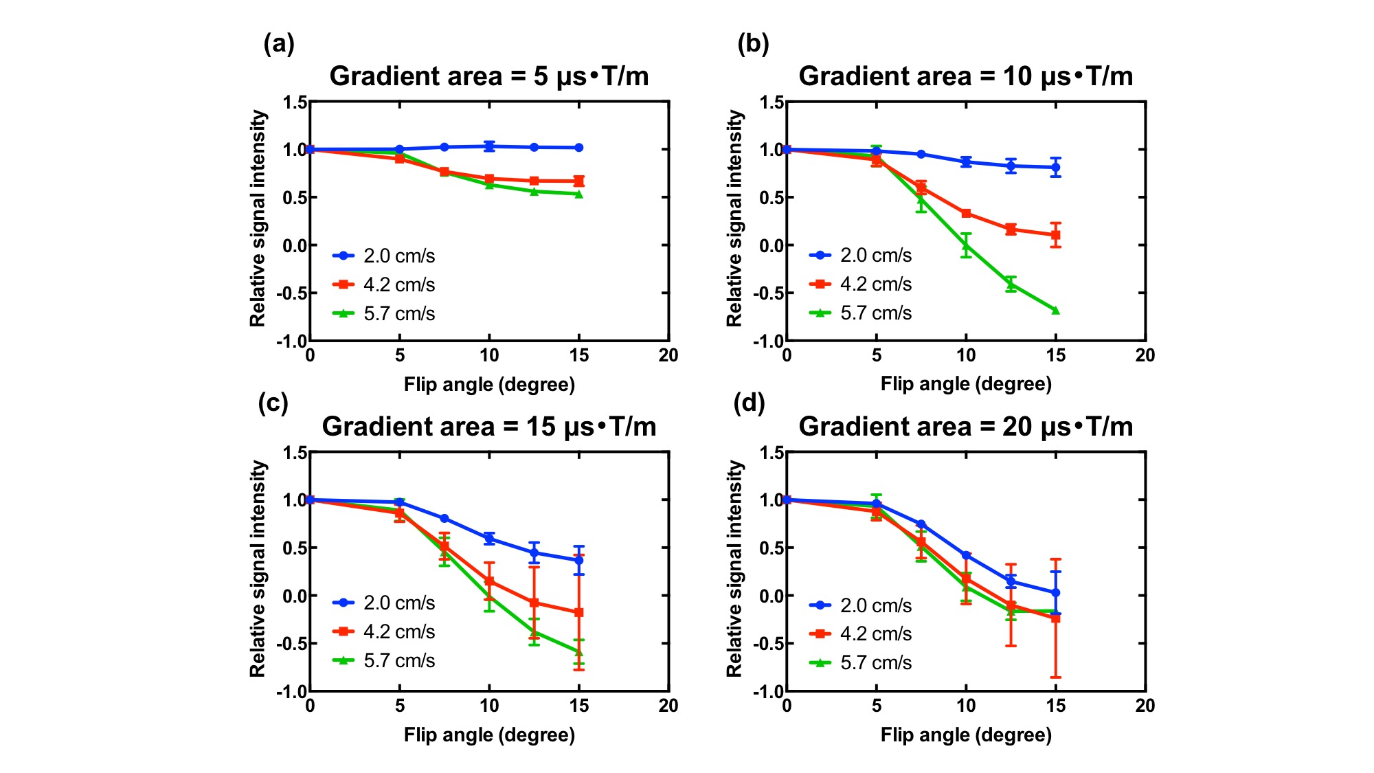
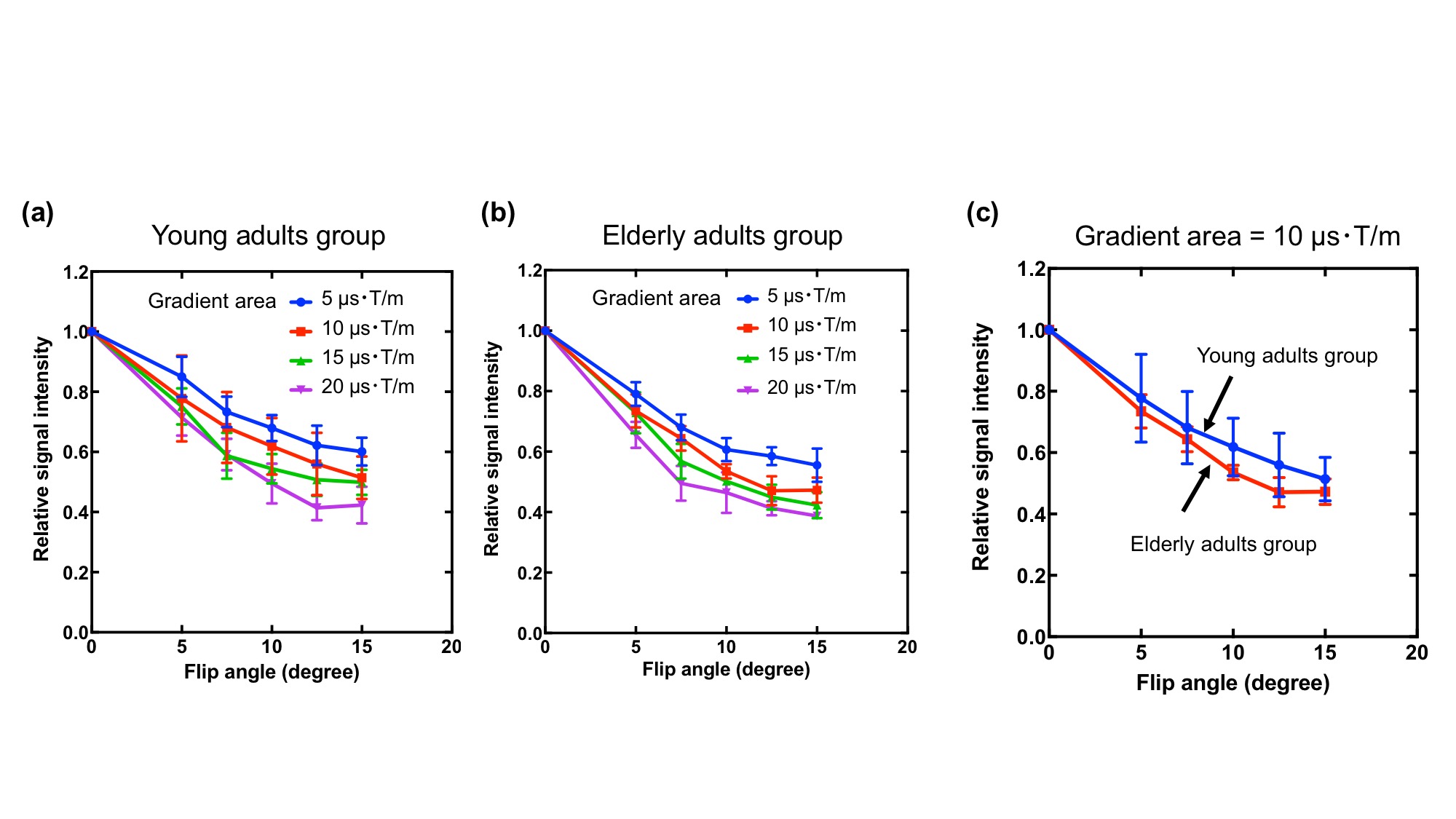
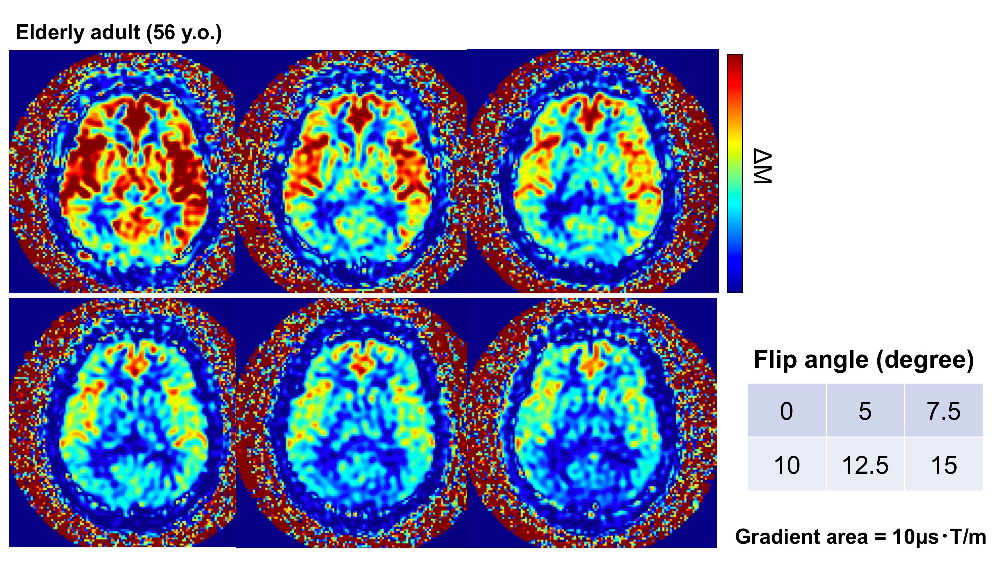
The results of a numerical simulation revealed that the magnetization of moving spins decreased in larger FA. The decrease of the magnetization was plateaued at FA = 12.5 degrees. On the other hand, the phantom study showed that the setting of larger FA or GA decreased the ASL signals (Fig. 2). The decrease of the ASL signal depends on the flow velocity, and the dependence increased with the decreasing GA. When the value of GA exceeded 15 μs・T/m, the standard deviation of the ASL signals increased. Based on the results of the in vivo study, larger FA and GA decreased the ΔM (Fig. 3). However, when GA > 15 μs・T/m and FA > 12.5 degrees, dispersion of the ΔM increased. Although there was no difference in the signal reduction in each vascular region, the ratio of signal decrease in elderly adult group were higher than that in young adult group (Fig. 4).
DISCUSSION AND CONCLUSION
The results of the phantom study showed that an optimal setting of the DANTE could make effectively decrease the signals in the vascular of a target flow velocity. In the in vivo study, the setting of larger GA and FA decreased the image uniformity or signal-to-noise ratio owing to the B1 inhomogeneity and eddy current, respectively. The decrease of ΔM by the DANTE pulse of the elderly adult groups was larger than that of young adult groups because the flow velocity of the elderly adults is lower than that of young adults. Therefore, it was concluded that an optimized DANTE pulse made it possible to efficiently and uniformly suppress the microvascular signal depending on the flow velocity. Moreover, the relative flow velocity map of labeled spins may be useful to assess altered hemodynamic state.Acknowledgements
No acknowledgement found.References
- Ye FQ, et al. Correction for vascular artifacts in cerebral blood flow values measured by using arterial spin tagging techniques. Magn. Reson. Med. 1997;37:226–235.
- Morris GA, Freeman R. Selective excitation in Fourier transform nuclear magnetic resonance. J Magn Reson 1978;29:433–462.
- Mosher TJ, Smith MB. A DANTE tagging sequence for the evaluation of translational sample motion. Magn Reson Med 1990;15:334–339.
- Li L, et al. DANTE-prepared pulse trains: A novel approach to motion-sensitized and motion-suppressed quantitative magnetic resonance imaging. Magn. Reson. Med. 2012;68:1423–1438.
Figures