1882
One minute Brain MR venography with Compressed SENSE at 3T.1Department of Diagnostic Imaging and Nuclear Medicine, Tokyo Women's Medical University, Tokyo, Japan
Synopsis
Brain MR venograpy based on phase-contrast technique (MRV) contributes in the diagnosis of venous sinus thrombosis and helps to clarify venous anatomy before brain operations. However, MRV is not commonly taken in routine brain MRI examinations because it requires a longer acquisition time. Recently, Compressed SENSE, which is a combination of compressed sensing and parallel imaging technique: SENSE, has been developed, and can shorten acquisition times with minimum image quality deterioration. Therefore, we investigated the optimization of 1 minute MRV, which the acquisition time was 1 minute, using Compressed SENSE at 3T.
INTRODUCTION
Brain MR venography based on phase-contrast technique (MRV) plays an important role to diagnose venous sinus thrombosis. 1, 2 Moreover, it is helpful to clarify venous anatomy before brain surgeries because the intracranial venous system has considerable anatomic variations.3, 4 However, MRV is not usually taken in a routine brain examination because MRV requires longer acquisition times and venous sinus thrombosis is an uncommon disease. Recently, Compressed SENSE (CS-SENSE), which is a combination of compressed sensing and parallel imaging technique: SENSE, has been developed to shorten acquisition times with minimum image quality deterioration. In this study, we evaluated the optimization of 1 minute MRV with CS-SENSE, which the acquisition time was 1 minute at 3T.METHODS
Five healthy volunteers were examined. (males: 3, females: 2, ages: 26 to 38 years) All studies were performed with 3T MR unit. (Ingenia 3T, Philips Healthcare R5.1.7) The different parameters between conventional MRV and 1 minute MRV with CS-SENSE is shown in Table 1. The other parameters were set as the same. (flow velocity: 10 cm/s.) Visualization of cerebral venous sinuses (Superior sagittal sinus: SSS, Transvers sinus: TS, Sigmoid sinus: SS, and Internal jugular vein: IJV) on conventional MRV and 1 minute MRV with CS-SENSE was evaluated.RESULTS
Cerebral venous sinuses: SSS, TS, SS, and IJV on 1 minute MRV with CS-SENSE were clearly visualized in all subjects, even though an increase in background noise was identified, compared with conventional MRV. Visualization of cortical veins on 1 minute MRV with CS-SENSE was slightly worse than with conventional MRV.DISCUSSION
MRV is expected to be excellent with CS-SENSE technique because signals of MRV have high sparsity. However, using an excessive high CS-SENSE factor causes an increase in background noise and signal loss of veins. In this study, setting both a suitable CS-SENSE factor and voxel size was very effective in shortening the acquisition time while keeping a high quality image resolution of cerebral venous sinuses. We suggest that an increase in background noise could lead to a slightly worse demonstration of cortical veins on 1 minute MRV with CS-SENSE. However, that degree would be no problem with the diagnosis of venous sinus thrombosis, because it is reported that it’s more important to detect signal change of cerebral venous sinuses than cortical veins, since cortical veins have more anatomical variations.2CONCLUSION
One minute MRV with CS-SENSE can provide excellent quality images equal to conventional MRV, and could be an informative technique for clinical use.Acknowledgements
No acknowledgement found.References
1. Loubeyre P, De Jaegere T, Tran-Minh VA. Three-dimensional phase contrast MR cerebral venography with zero filling interpolation in the slice encoding direction. Magn Reson Imaging. 1999;17(8):1227-1233.
2. Sajjad Z. MRI and MRV in cerebral venous thrombosis. J Pak Med Assoc. 2006;56(11):523-526.
3. Ayanzen RH, Bird CR, Keller PJ, et al. Cerebral venography: normal anatomy and potential diagnostic pitfalls. AJNR Am J Neuroradiol 2000;21:74-78
4. Goyal G, Singh R, Bansal N, et al. Anatomical variations of cerebral MR venography: is gender matter? Neurointervention. 2016;11(2):92-98
Figures
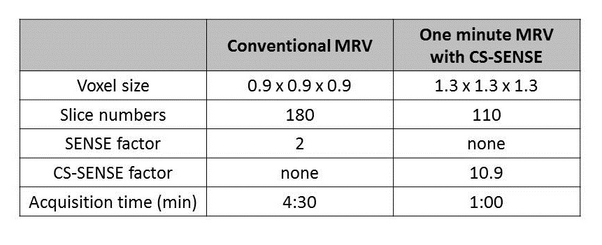
Table 1. The different imaging parameters of conventional MRV and 1 minute MRV with CS-SENSE.
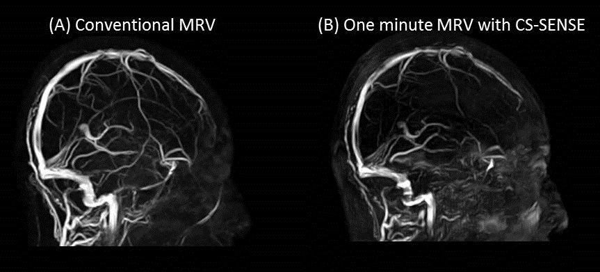
Figure 1. Images on conventional MRV and 1 minute MRV with CS-SENSE.
(A) Conventional MRV (B) One minute MRV with CS-SENSE
Cerebral venous sinuses (Superior sagittal sinus, Transvers sinus, Sigmoid sinus, and Internal jugular vein) are clearly visualized on both conventional MRV and 1 minute MRV with CS-SENSE. One minute MRV with CS-SENSE shows an increase in background noise and slightly less visualization of cortical veins.