1880
Template maps of vascular function and structure in the healthy brain1Department for Diagnostic Physics, Oslo University Hospital, Oslo, Norway, 2Athinoula A. Martinos Center for Biomedical Imaging, Harvard Medical School, Boston, MA, United States
Synopsis
In recent years, Vessel Architectural Imaging (VAI) has emerged as a promising tool in tumor diagnosis to reveal unique MRI-based information on vessel architecture, hemodynamic efficacy and metabolic activity. Healthy control data may further advance our knowledge on the VAI method and its underlying mechanisms, as well as serve as study controls. Here we propose a set of healthy-tissue template maps of all VAI derived parameters which may act as a toolbox to identify anomalies of various vascular brain diseases and ultimately help improve diagnostic and outcome assessment in clinical settings.
INTRODUCTION
Vessel Architectural Imaging (VAI) has recently been introduced as a new paradigm in tumor diagnosis (1). The VAI technique exploits a temporal shift in the vessel-size sensitive MRI signals to revel new information on vessel calibers, architecture, hemodynamic efficacy and metabolic activity (2). Here, we propose a set of templates for VAI parameters derived from MRIs of volunteers, thus serving as a potential control to identify anomalies of vascular brain diseases.MATERIALS AND METHODS
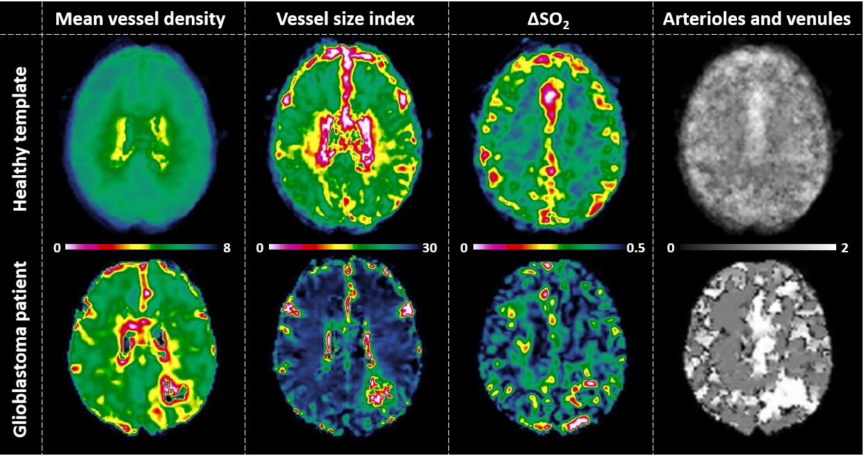
In this IRB-approved study, ten healthy volunteers underwent contrast-enhanced MRI (Skyra 3T, Siemens) including gradient-echo spin-echo dynamic susceptibility contrast (DSC) imaging with simultaneous-multiple-slice (SMS) acceleration (3). Parametric maps of mean vessel density, vessel size index, oxygenation status (∆SO2), and voxel-wise dominance of arterioles and venules was estimated by VAI as previously described (1) and used as proof-of-concept hereafter. The resulting parametrical maps were spatially normalized in MNI-space using the SPM software package. For all normalized maps, template maps were generated by estimating the mean of all 10 healthy subjects. In addition, one glioblastoma patient was included as a case example for comparison.RESULTS
This proof-of-concept study show the initial framework of healthy-tissue template maps of vascular function in the brain. A total of four template maps were generated based on the healthy subjects, as shown in Figure 1. This figure also shows the comparison to a glioblastoma patient, in which substantial differences in vascular function are shown, thus demonstrating the potential clinical value.CONCLUSION
We propose a set of template maps of healthy vascular function of the brain that may serve as a toolbox for early identification of anomalies of vascular brain disease, with the ultimate goal of improving the treatment decision making.Acknowledgements
No acknowledgement found.References
1. Emblem KE et al. Nat Med. 2013;19(9):1178-1183 (doi: 10.1038/nm.3289)
2. Digernes I et al. J Cereb Blood Flow Metab. 2017;37(6):2237-2248 (doi: 10.1177/0271678X17694187)
3. Eichner C et al. Magn Reson Med. 2014;72(3):770-778 (doi: 10.1002/mrm.24960)
Figures