1879
Vascular Change Assessed by Calibrated Multi-delay Arterial Spin Labeling Under Oxygen and Carbogen Gas ChallengeMichael L Rohan1, Clara Wellons2, Megan Shevenell3, Nicolette Schwarz4, Xingfeng Shao5, Daniel JJ Wang5, and Blaise Frederick3
1Imaging, McLean Hospital, Belmont, MA, United States, 2imaging, McLean Hospital, Belmont, MA, United States, 3McLean Hospital, Belmont, MA, United States, 4Mc:ean Hospital, Belmont, MA, United States, 5University of Southern California, Los Angeles, CA, United States
Synopsis
Arterial Spin Labeling (ASL) measurements are employed here in a suite of hemodynamic assessments in our study of cerebrovascular reactivity. In this project we test a 3D Gradient and Spin Echo (GRASE) Multiple delay Pseudo-Continuous Arterial Spin Labeling (MPCASL) Magnetic Resonance (MR) acquisition in order to measure the change in these measures under gas challenge. Subjects were scanned with 3D GRASE MPCASL while breathing medical air, oxygen, or Carbogen (5% CO2 +95% O2) under controlled conditions. Changes in blood flow, volume, and arrival time that were observed will be used to calibrate novel delay assessment methods.
Introduction
Brain health requires that the cerebral vasculature be able to supply an appropriate amount of oxygen and nutrients to brain tissue under a wide range of physiological conditions. These conditions include both the normal resting baseline state, and states with higher oxygen requirements, such as high cognitive load; to meet this requirement, the vasculature undergoes autoregulatory vasodilation to respond to increase demand. Cerebrovascular reactivity (CVR) is the brain blood vessels’ capacity for vasodilation. It offers useful clinical information in patients at risk for cerebral ischemia associated with chronic stenosis or occlusion of cerebral blood vessels, patients at risk of ischemia in deep white matter, which leads to sub-cortical infarction, and may reflect abnormalities in diabetes, and in patients with dementing illnesses including Alzheimer’s disease. Assessment of hemodynamic impairment may be critical to informing management decisions in patients with atherosclerotic disease, and neuroimaging is performed routinely in the evaluation of these patients. This project will calibrate our MPCASL sequence and methodology using Oxygen and Carbon dioxide gas challenges to simulate vascular pathology. Arterial Spin Labeling is an MRI technique used to study cerebral blood flow and hemodynamics. In this project we use static ASL to assess hemodynamic parameters such as cerebral blood flow (CBF), cerebral blood volume (CBV), and bolus arrival time. In order to simulate pathology we perform these measures in healthy controls while breathing medical air (MA), Oxygen (100% O2), or Carbogen (5% CO2 / 95% O2). Oxygen is a vasoconstrictor and carbon dioxide is a vasodilator; and we expected both increases and decreases in hemodynamic parameters to occur under these gas challenges. We expect O2 to have minimal effects on CBF and CBV; we expect Carbogen to cause increases in CBF and in bolus arrival time.Methods
We collected multi-delay PCASL data using a 3D Gradient and Spin Echo (GRASE) sequence on a Siemens 3T Prisma MR system. 3D GRASE provides higher resolution within achievable scan times. The acquisition used 8 single volume delays with TE=30ms, TR=4s, TI = 1.6s, 2.0s, 2.4s, 2.8s, 3.2s, 3.6s, 4.0s and 4.4s; the tag duration was 1.5s (within TI); isotropic voxels were 3.5mm with a matrix of 64x64x40; scan time was 5:30. Three acquisitions were made. Subjects were breathing either MA, O2 or Carbogen through an MR compatible portable tank-based apparatus utilizing a demand regulator during each scan. The sequence provided both tag and control images as well as M0 images. A standard 3D anatomic image was also acquired for registration. Data were acquired from five healthy subjects. Calibrated ASL processing was performed using FSL BASIL software (OSFORD FMRIB v5.0.10) which provided maps of voxel wise bolus arrival time, quantitative CBF and quantitative CBV. Assessments were made of the three hemodynamic parameters in a gray matter (GM) region and in a region containing the basal ganglia and thalamus (BG). These two regions were selected because they represent different tissue vasculature and variability for comparison. Regional mean values were compared for CBF and CBV, while voxel wise maps of the relative change in arrival time from MA to O2 and MA to Carbogen were made to assess change in arrival time.Results
Changes in CBF were apparent with a reduction in regional CBF observed under O2 as compared to MA, 18.5% in both GM and the BG; there was an increase in CBF under Carbogen in GM (16.5%) and in the BG (6%). CBV increased under the influence of both gases, with a 13.8% increase under O2 in GM and a 27.8% increase under O2 in the BG, and an increase by 26.8% under Carbogen in the GM and by 45% in the BG. Arrival times were unchanged in both regions under O2 and were decreased by 3% in the GM and by 2% in the BG under Carbogen. The regional mean changes are shown in the figures below (CBF and CBV) as well as the mean voxel wise change in bolus arrival times.Discussion
The repeatability and robustness of these results demonstrate that the 3D GRASE sequence can be used to detect the desired changes in cerebrovascular reactivity. Reduction in CBF under the influence of O2 occurred as expected, and the increase in CBF under the influence of the carbon dioxide in the Carbogen mix was also as expected. The increase in CBV under both gases was not expected and further modeling will be performed to explain this. The changes in bolus arrival time were small but robust.Acknowledgements
No acknowledgement found.References
Donahue, M.J., et al., Bolus arrival time and cerebral blood flow responses to hypercarbia. Journal of cerebral blood flow and metabolism : official journal of the International Society of Cerebral Blood Flow and Metabolism, 2014. 34(7): p. 1243-1252. Alsop, D.C., et al., Recommended implementation of arterial spin-labeled perfusion MRI for clinical applications: A consensus of the ISMRM perfusion study group and the European consortium for ASL in dementia. Magnetic resonance in medicine : official journal of the Society of Magnetic Resonance in Medicine / Society of Magnetic Resonance in Medicine, 2014. Wang et al. Neuroimage Clin. 2013;3:1-7Figures
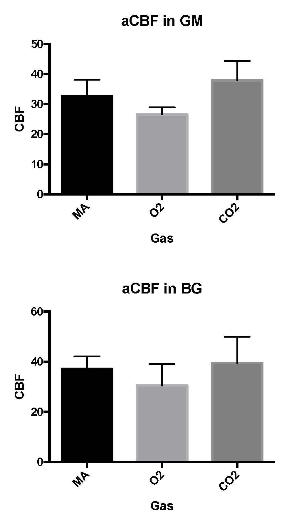
CBF
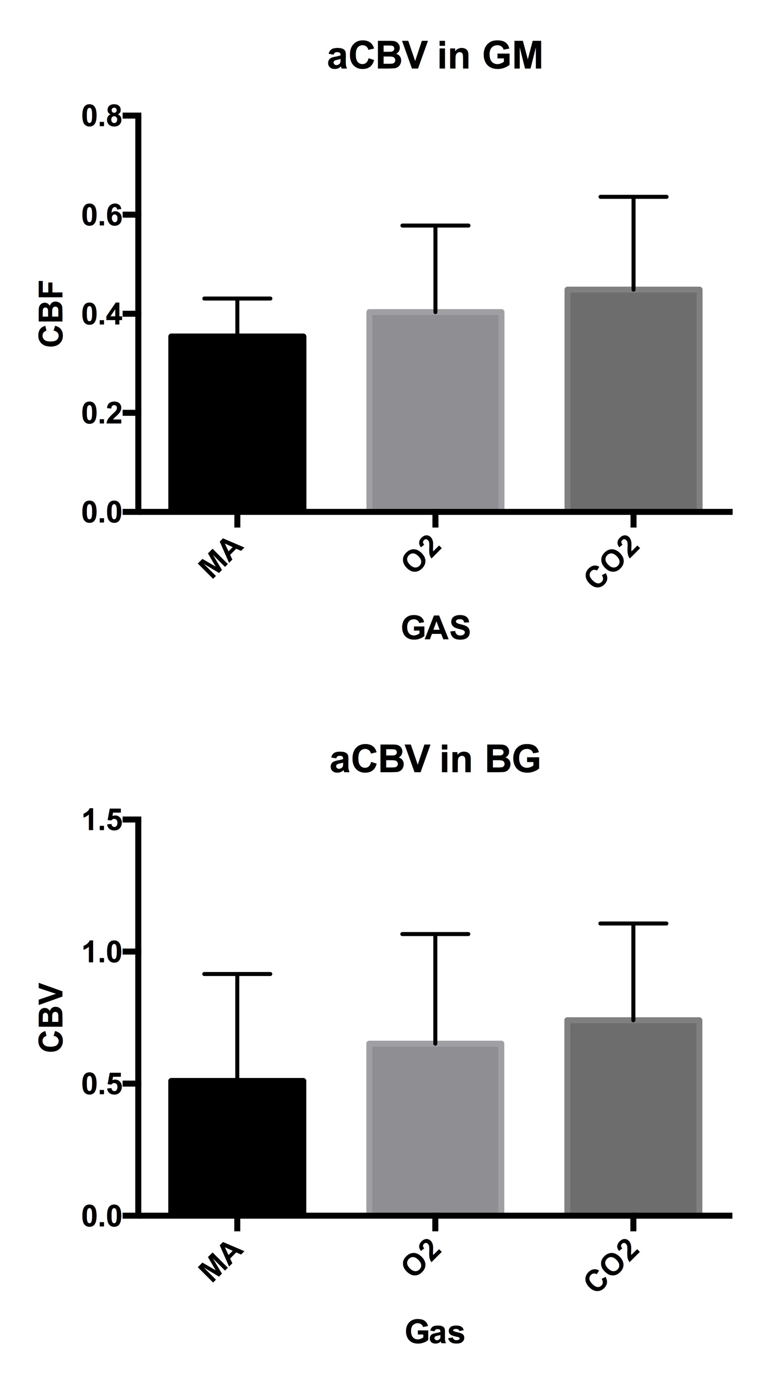
CBV
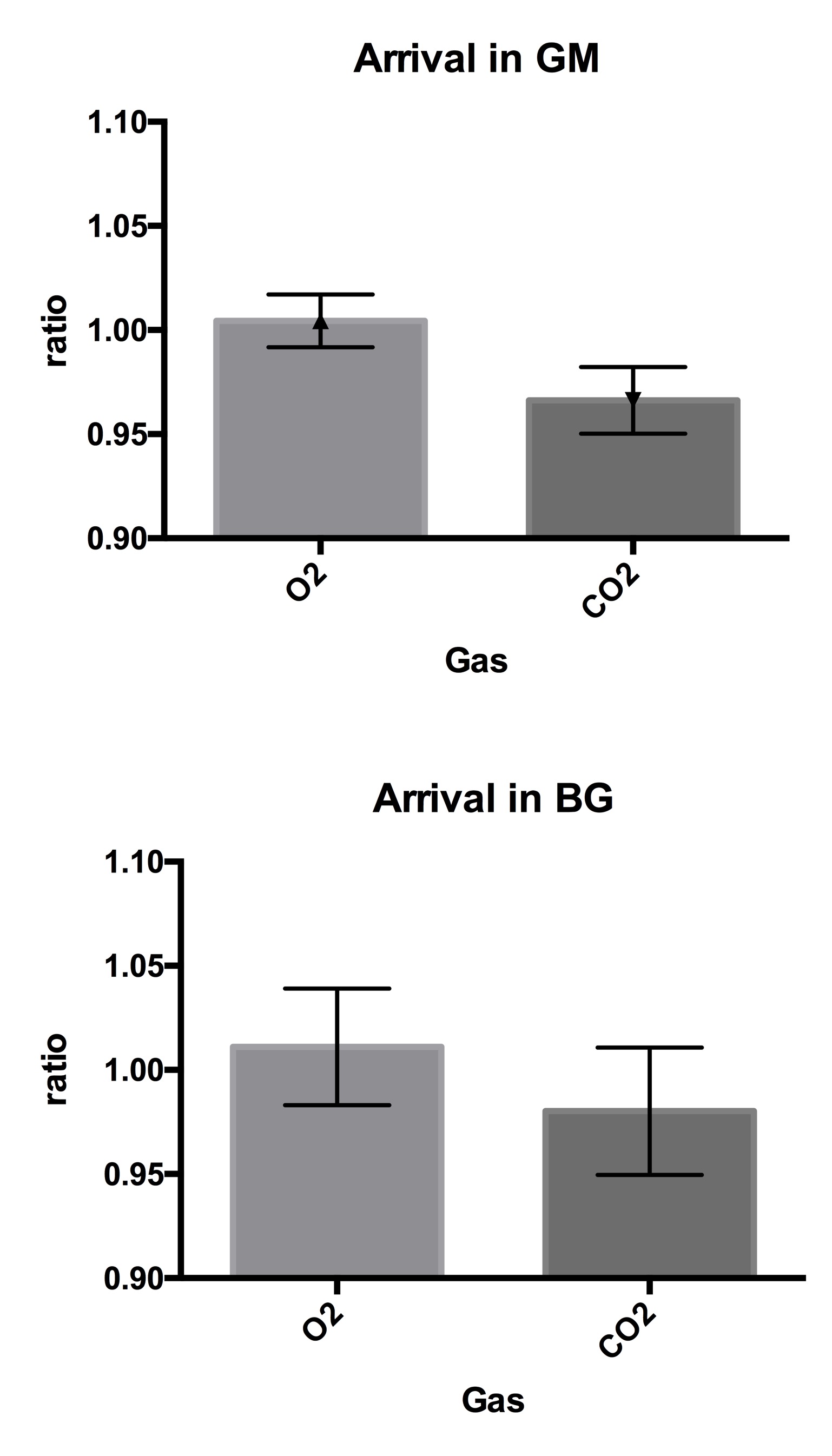
Arrival